 Movie
Movie Controller

[English] 日本語
 Yorodumi
- PDB-7q19: Beta-lactoglobulin mutant FAW (I56F/L39A/M107W) in complex with d... -
+ Open data

- Basic information
| Entry | Database: PDB / ID: 7q19 | ||||||
|---|---|---|---|---|---|---|---|
| Title | Beta-lactoglobulin mutant FAW (I56F/L39A/M107W) in complex with desipramine (FAW-DSM#3) | ||||||
 Components Components | Beta-lactoglobulin | ||||||
 Keywords Keywords | TRANSPORT PROTEIN / lactoglobulin / mutation / desipramine | ||||||
| Function / homology |  Function and homology information Function and homology informationretinol binding / long-chain fatty acid binding / extracellular region / identical protein binding Similarity search - Function | ||||||
| Biological species |  | ||||||
| Method |  X-RAY DIFFRACTION / SYNCHROTRON / MOLECULAR REPLACEMENT / Resolution: 1.55 Å X-RAY DIFFRACTION / SYNCHROTRON / MOLECULAR REPLACEMENT / Resolution: 1.55 Å | ||||||
 Authors Authors | Loch, J.I. / Barciszewski, J. / Pokrywka, K. / Lewinski, K. | ||||||
| Funding support | 1items
| ||||||
 Citation Citation | Journal: Iucrj / Year: 2022 Title: New ligand-binding sites identified in the crystal structures of [beta]-lactoglobulin complexes with desipramine Authors: Loch, J.I. / Barciszewski, J. / Sliwiak, J. / Bonarek, P. / Wrobel, P. / Pokrywka, K. / Shabalin, I.G. / Minor, W. / Jaskolski, M. / Lewinski, K. | ||||||
| History |
|
- Structure visualization
| Structure viewer | Molecule: MolmilJmol/JSmol |
|---|
- Downloads & links
-Download
| PDBx/mmCIF format | 7q19.cif.gz | 78.2 KB | Display | PDBx/mmCIF format |
|---|---|---|---|---|
| PDB format | pdb7q19.ent.gz | Display | PDB format | |
| PDBx/mmJSON format | 7q19.json.gz | Tree view | PDBx/mmJSON format | |
| Others |  Other downloads Other downloads |
-Validation report
| Arichive directory | https://data.pdbj.org/pub/pdb/validation_reports/q1/7q19ftp://data.pdbj.org/pub/pdb/validation_reports/q1/7q19 | HTTPS FTP |
|---|
-Related structure data
| Related structure data |  7q17C 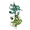 7q18C  7q2nC  7q2oC  7q2pC 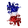 1bsyS S: Starting model for refinement C: citing same article ( |
|---|---|
| Similar structure data |
-Links
 PDBj
PDBj

- Assembly
| Deposited unit | 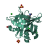
| |||||||||||||||||||||
|---|---|---|---|---|---|---|---|---|---|---|---|---|---|---|---|---|---|---|---|---|---|---|
| 1 | 
| |||||||||||||||||||||
| Unit cell |
| |||||||||||||||||||||
| Components on special symmetry positions |
|
-Components
-Protein , 1 types, 1 molecules AAA
| #1: Protein | Mass: 18279.965 Da / Num. of mol.: 1 / Mutation: L1A, I2S, L39A, I56F, M107W Source method: isolated from a genetically manipulated source Source: (gene. exp.)  |
|---|
-Non-polymers , 5 types, 127 molecules 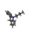


| #2: Chemical | ChemComp-DSM /  Mass: 266.381 Da / Num. of mol.: 1 / Source method: obtained synthetically / Formula: C18H22N2 / Feature type: SUBJECT OF INVESTIGATION / Comment: antidepressant, inhibitor*YM Mass: 266.381 Da / Num. of mol.: 1 / Source method: obtained synthetically / Formula: C18H22N2 / Feature type: SUBJECT OF INVESTIGATION / Comment: antidepressant, inhibitor*YM | ||||
|---|---|---|---|---|---|
| #3: Chemical | ChemComp-EDO /  Mass: 62.068 Da / Num. of mol.: 1 / Source method: obtained synthetically / Formula: C2H6O2 Mass: 62.068 Da / Num. of mol.: 1 / Source method: obtained synthetically / Formula: C2H6O2 | ||||
| #4: Chemical |  Mass: 96.063 Da / Num. of mol.: 2 / Source method: obtained synthetically / Formula: SO4 Mass: 96.063 Da / Num. of mol.: 2 / Source method: obtained synthetically / Formula: SO4#5: Chemical |  Mass: 35.453 Da / Num. of mol.: 3 / Source method: obtained synthetically / Formula: Cl Mass: 35.453 Da / Num. of mol.: 3 / Source method: obtained synthetically / Formula: Cl#6: Water | ChemComp-HOH / | Mass: 18.015 Da / Num. of mol.: 120 / Source method: isolated from a natural source / Formula: H2O |
-Details
| Has ligand of interest | Y |
|---|---|
| Has protein modification | Y |
-Experimental details
-Experiment
| Experiment | Method: X-RAY DIFFRACTION / Number of used crystals: 1 |
|---|
- Sample preparation
| Crystal | Density Matthews: 2.1 Å3/Da / Density % sol: 41.55 % |
|---|---|
| Crystal grow | Temperature: 293 K / Method: vapor diffusion, hanging drop / pH: 8.5 Details: 2.40 M (NH4)2SO4, 0.5 M Tris-HCl pH 8.5, desipramine |
-Data collection
| Diffraction | Mean temperature: 100 K / Serial crystal experiment: N |
|---|---|
| Diffraction source | Source: SYNCHROTRON / Site: BESSY  / Beamline: 14.1 / Wavelength: 0.9184 Å / Beamline: 14.1 / Wavelength: 0.9184 Å |
| Detector | Type: DECTRIS PILATUS3 6M / Detector: PIXEL / Date: Apr 23, 2019 |
| Radiation | Protocol: SINGLE WAVELENGTH / Monochromatic (M) / Laue (L): M / Scattering type: x-ray |
| Radiation wavelength | Wavelength: 0.9184 Å / Relative weight: 1 |
| Reflection | Resolution: 1.55→41.69 Å / Num. obs: 22796 / % possible obs: 99.9 % / Redundancy: 10.3 % / CC1/2: 1 / Rrim(I) all: 0.051 / Net I/σ(I): 23.24 |
| Reflection shell | Resolution: 1.55→1.64 Å / Redundancy: 9.11 % / Mean I/σ(I) obs: 1.98 / Num. unique obs: 3482 / CC1/2: 0.087 / Rrim(I) all: 1.022 / % possible all: 99.4 |
- Processing
| Software |
| |||||||||||||||||||||||||||||||||||||||||||||||||||||||||||||||||||||||||||||||||||||||||||||||||||||||||||||||||||||||||||||||||||||||||||||||||||||||||||
|---|---|---|---|---|---|---|---|---|---|---|---|---|---|---|---|---|---|---|---|---|---|---|---|---|---|---|---|---|---|---|---|---|---|---|---|---|---|---|---|---|---|---|---|---|---|---|---|---|---|---|---|---|---|---|---|---|---|---|---|---|---|---|---|---|---|---|---|---|---|---|---|---|---|---|---|---|---|---|---|---|---|---|---|---|---|---|---|---|---|---|---|---|---|---|---|---|---|---|---|---|---|---|---|---|---|---|---|---|---|---|---|---|---|---|---|---|---|---|---|---|---|---|---|---|---|---|---|---|---|---|---|---|---|---|---|---|---|---|---|---|---|---|---|---|---|---|---|---|---|---|---|---|---|---|---|---|
| Refinement | Method to determine structure: MOLECULAR REPLACEMENT Starting model: 1BSY Resolution: 1.55→41.69 Å / Cor.coef. Fo:Fc: 0.965 / Cor.coef. Fo:Fc free: 0.954 / SU B: 4.015 / SU ML: 0.069 / Cross valid method: FREE R-VALUE / ESU R: 0.092 / ESU R Free: 0.094
| |||||||||||||||||||||||||||||||||||||||||||||||||||||||||||||||||||||||||||||||||||||||||||||||||||||||||||||||||||||||||||||||||||||||||||||||||||||||||||
| Solvent computation | Ion probe radii: 0.8 Å / Shrinkage radii: 0.8 Å / VDW probe radii: 1.2 Å / Solvent model: MASK BULK SOLVENT | |||||||||||||||||||||||||||||||||||||||||||||||||||||||||||||||||||||||||||||||||||||||||||||||||||||||||||||||||||||||||||||||||||||||||||||||||||||||||||
| Displacement parameters | Biso mean: 29.44 Å2
| |||||||||||||||||||||||||||||||||||||||||||||||||||||||||||||||||||||||||||||||||||||||||||||||||||||||||||||||||||||||||||||||||||||||||||||||||||||||||||
| Refinement step | Cycle: LAST / Resolution: 1.55→41.69 Å
| |||||||||||||||||||||||||||||||||||||||||||||||||||||||||||||||||||||||||||||||||||||||||||||||||||||||||||||||||||||||||||||||||||||||||||||||||||||||||||
| Refine LS restraints |
| |||||||||||||||||||||||||||||||||||||||||||||||||||||||||||||||||||||||||||||||||||||||||||||||||||||||||||||||||||||||||||||||||||||||||||||||||||||||||||
| LS refinement shell |
| |||||||||||||||||||||||||||||||||||||||||||||||||||||||||||||||||||||||||||||||||||||||||||||||||||||||||||||||||||||||||||||||||||||||||||||||||||||||||||
| Refinement TLS params. | Method: refined / Origin x: -12.8202 Å / Origin y: 52.1562 Å / Origin z: 15.2609 Å
| |||||||||||||||||||||||||||||||||||||||||||||||||||||||||||||||||||||||||||||||||||||||||||||||||||||||||||||||||||||||||||||||||||||||||||||||||||||||||||
| Refinement TLS group |
|
