 Movie
Movie Controller


+ Open data

- Basic information
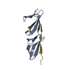
| Entry | Database: PDB / ID: 7o39 | ||||||
|---|---|---|---|---|---|---|---|
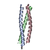
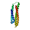
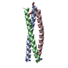
| Title | Crystal structure of S. aureus DivIVA N terminal domain | ||||||
 Components Components | DivIVA | ||||||
 Keywords Keywords | PROTEIN BINDING / Cell division / Divisome / molecular ruler | ||||||
| Function / homology | DivIVA family / DivIVA domain / DivIVA protein / cytoplasm / DivIVA domain-containing protein Function and homology information Function and homology information | ||||||
| Biological species |   Staphylococcus aureus (bacteria) Staphylococcus aureus (bacteria) | ||||||
| Method |  X-RAY DIFFRACTION / SYNCHROTRON / MOLECULAR REPLACEMENT / Resolution: 1.3 Å X-RAY DIFFRACTION / SYNCHROTRON / MOLECULAR REPLACEMENT / Resolution: 1.3 Å | ||||||
 Authors Authors | Rao, V.A. / Booth, S. / Lewis, R.J. | ||||||
| Funding support |  United Kingdom, 1items United Kingdom, 1items
| ||||||
 Citation Citation | Journal: To Be Published Title: Crystal structure of S. aureus DivIVA N terminal domain Authors: Rao, V.A. / Booth, S. / Lewis, R.J. | ||||||
| History |
|
- Structure visualization
| Structure viewer | Molecule: MolmilJmol/JSmol |
|---|
- Downloads & links
-Download
| PDBx/mmCIF format | 7o39.cif.gz | 90.5 KB | Display | PDBx/mmCIF format |
|---|---|---|---|---|
| PDB format | pdb7o39.ent.gz | 63.8 KB | Display | PDB format |
| PDBx/mmJSON format | 7o39.json.gz | Tree view | PDBx/mmJSON format | |
| Others |  Other downloads Other downloads |
-Validation report
| Arichive directory | https://data.pdbj.org/pub/pdb/validation_reports/o3/7o39ftp://data.pdbj.org/pub/pdb/validation_reports/o3/7o39 | HTTPS FTP |
|---|
-Related structure data
| Related structure data | 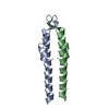 2wujS S: Starting model for refinement |
|---|---|
| Similar structure data |


-Links
 PDBj
PDBj- Assembly
| Deposited unit | 
| ||||||||||||
|---|---|---|---|---|---|---|---|---|---|---|---|---|---|
| 1 |
| ||||||||||||
| Unit cell |
|
-Components
| #1: Protein | Mass: 6909.777 Da / Num. of mol.: 2 Source method: isolated from a genetically manipulated source Source: (gene. exp.) Staphylococcus aureus (strain NCTC 8325 / PS 47) (bacteria)Strain: NCTC 8325 / PS 47 / Gene: SAOUHSC_01158 / Plasmid: pOPINRSF / Production host: #2: Water | ChemComp-HOH / |  Mass: 18.015 Da / Num. of mol.: 172 / Source method: isolated from a natural source / Formula: H2O Mass: 18.015 Da / Num. of mol.: 172 / Source method: isolated from a natural source / Formula: H2O |
|---|
-Experimental details
-Experiment
| Experiment | Method: X-RAY DIFFRACTION / Number of used crystals: 1 |
|---|
- Sample preparation
| Crystal | Density Matthews: 2.22 Å3/Da / Density % sol: 44.62 % / Description: Cubic |
|---|---|
| Crystal grow | Temperature: 293 K / Method: vapor diffusion, sitting drop / pH: 6.5 Details: 0.1 M Carboxylic acids, 0.1 M Buffer system 1 pH 6.5, 37.5 % v/v MPD_P1K_P3350; Morpheus G4 |
-Data collection
| Diffraction | Mean temperature: 100 K / Serial crystal experiment: N |
|---|---|
| Diffraction source | Source: SYNCHROTRON / Site: Diamond / Beamline: I03 / Wavelength: 0.97623 Å |
| Detector | Type: DECTRIS PILATUS3 6M / Detector: PIXEL / Date: Feb 5, 2018 |
| Radiation | Protocol: SINGLE WAVELENGTH / Monochromatic (M) / Laue (L): M / Scattering type: x-ray |
| Radiation wavelength | Wavelength: 0.97623 Å / Relative weight: 1 |
| Reflection | Resolution: 1.3→37.08 Å / Num. obs: 31216 / % possible obs: 100 % / Redundancy: 6.9 % / Biso Wilson estimate: 15.87 Å2 / CC1/2: 0.99 / Rmerge(I) obs: 0.087 / Rpim(I) all: 0.051 / Rrim(I) all: 0.101 / Χ2: 0.8 / Net I/σ(I): 10.9 |
| Reflection shell | Resolution: 1.3→1.32 Å / Redundancy: 6.8 % / Rmerge(I) obs: 0.556 / Mean I/σ(I) obs: 2.5 / Num. unique obs: 1504 / CC1/2: 0.904 / Rpim(I) all: 0.339 / Rrim(I) all: 0.653 / Χ2: 0.33 / % possible all: 99.6 |
- Processing
| Software |
| ||||||||||||||||||||||||||||||||||||||||||||||||||||||||||||||||||||||||||||||||||||
|---|---|---|---|---|---|---|---|---|---|---|---|---|---|---|---|---|---|---|---|---|---|---|---|---|---|---|---|---|---|---|---|---|---|---|---|---|---|---|---|---|---|---|---|---|---|---|---|---|---|---|---|---|---|---|---|---|---|---|---|---|---|---|---|---|---|---|---|---|---|---|---|---|---|---|---|---|---|---|---|---|---|---|---|---|---|
| Refinement | Method to determine structure: MOLECULAR REPLACEMENT Starting model: 2WUJ Resolution: 1.3→36.95 Å / SU ML: 0.1175 / Cross valid method: FREE R-VALUE / σ(F): 1.38 / Phase error: 15.2283 Stereochemistry target values: GeoStd + Monomer Library + CDL v1.2
| ||||||||||||||||||||||||||||||||||||||||||||||||||||||||||||||||||||||||||||||||||||
| Solvent computation | Shrinkage radii: 0.9 Å / VDW probe radii: 1.11 Å / Solvent model: FLAT BULK SOLVENT MODEL | ||||||||||||||||||||||||||||||||||||||||||||||||||||||||||||||||||||||||||||||||||||
| Displacement parameters | Biso mean: 23.98 Å2 | ||||||||||||||||||||||||||||||||||||||||||||||||||||||||||||||||||||||||||||||||||||
| Refinement step | Cycle: LAST / Resolution: 1.3→36.95 Å
| ||||||||||||||||||||||||||||||||||||||||||||||||||||||||||||||||||||||||||||||||||||
| Refine LS restraints |
| ||||||||||||||||||||||||||||||||||||||||||||||||||||||||||||||||||||||||||||||||||||
| LS refinement shell |
|
