 Movie
Movie Controller

[English] 日本語
 Yorodumi
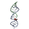
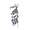
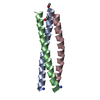
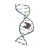
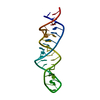
- PDB-5vci: RNA hairpin structure containing tetraloop/receptor motif, comple... -
+ Open data

- Basic information
| Entry | Database: PDB / ID: 5vci | ||||||
|---|---|---|---|---|---|---|---|
| Title | RNA hairpin structure containing tetraloop/receptor motif, complexed with 2-MeImpG analogue | ||||||
 Components Components |
| ||||||
 Keywords Keywords | RNA / hairpin / tetraloop/receptor | ||||||
| Function / homology | Chem-8OS / RNA / RNA (> 10) Function and homology information Function and homology information | ||||||
| Biological species | synthetic construct (others) | ||||||
| Method |  X-RAY DIFFRACTION / SYNCHROTRON / MOLECULAR REPLACEMENT / Resolution: 2.6 Å X-RAY DIFFRACTION / SYNCHROTRON / MOLECULAR REPLACEMENT / Resolution: 2.6 Å | ||||||
 Authors Authors | Szostak, J.W. / Zhang, W. | ||||||
 Citation Citation | Journal: J. Am. Chem. Soc. / Year: 2018 Title: Structural Rationale for the Enhanced Catalysis of Nonenzymatic RNA Primer Extension by a Downstream Oligonucleotide. Authors: Zhang, W. / Tam, C.P. / Zhou, L. / Oh, S.S. / Wang, J. / Szostak, J.W. | ||||||
| History |
|
- Structure visualization
| Structure viewer | Molecule: MolmilJmol/JSmol |
|---|
- Downloads & links
-Download
| PDBx/mmCIF format | 5vci.cif.gz | 31.1 KB | Display | PDBx/mmCIF format |
|---|---|---|---|---|
| PDB format | pdb5vci.ent.gz | 19.9 KB | Display | PDB format |
| PDBx/mmJSON format | 5vci.json.gz | Tree view | PDBx/mmJSON format | |
| Others |  Other downloads Other downloads |
-Validation report
| Arichive directory | https://data.pdbj.org/pub/pdb/validation_reports/vc/5vciftp://data.pdbj.org/pub/pdb/validation_reports/vc/5vci | HTTPS FTP |
|---|
-Related structure data
| Related structure data |  5ux3C 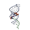 5uz6C 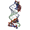 5v0hC 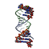 5v0jC 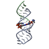 5v0oC 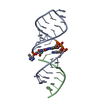 5v9zC  5vcfC  5vgwC 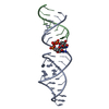 6az4C  6bmdC  4fnjS C: citing same article ( S: Starting model for refinement |
|---|---|
| Similar structure data |


-Links
 PDBj
PDBj


- Assembly
| Deposited unit | 
| ||||||||
|---|---|---|---|---|---|---|---|---|---|
| 1 |
| ||||||||
| Unit cell |
|
-Components
| #1: RNA chain | Mass: 7112.306 Da / Num. of mol.: 1 / Source method: obtained synthetically / Source: (synth.) synthetic construct (others) |
|---|---|
| #2: RNA chain | Mass: 3765.256 Da / Num. of mol.: 1 / Source method: obtained synthetically / Source: (synth.) synthetic construct (others) |
| #3: Chemical | ChemComp-8OS /   Type: RNA linking / Mass: 427.309 Da / Num. of mol.: 1 / Source method: obtained synthetically / Formula: C14H18N7O7P Type: RNA linking / Mass: 427.309 Da / Num. of mol.: 1 / Source method: obtained synthetically / Formula: C14H18N7O7P |
| #4: Water | ChemComp-HOH /  Mass: 18.015 Da / Num. of mol.: 1 / Source method: isolated from a natural source / Formula: H2O Mass: 18.015 Da / Num. of mol.: 1 / Source method: isolated from a natural source / Formula: H2O |
-Experimental details
-Experiment
| Experiment | Method: X-RAY DIFFRACTION / Number of used crystals: 1 |
|---|
- Sample preparation
| Crystal | Density Matthews: 3.2 Å3/Da / Density % sol: 61.5 % |
|---|---|
| Crystal grow | Temperature: 291 K / Method: vapor diffusion, sitting drop / pH: 7 Details: 50 mM MgCl2, 0.2 M Lithium sulfate monohydrate, 0.1 M BIS-TRIS pH 7.0, 25% w/v Polyethylene glycol 3,350 |
-Data collection
| Diffraction | Mean temperature: 99 K |
|---|---|
| Diffraction source | Source: SYNCHROTRON / Site: ALS  / Beamline: 8.2.2 / Wavelength: 1 Å / Beamline: 8.2.2 / Wavelength: 1 Å |
| Detector | Type: ADSC QUANTUM 315 / Detector: CCD / Date: Oct 28, 2016 |
| Radiation | Protocol: SINGLE WAVELENGTH / Monochromatic (M) / Laue (L): M / Scattering type: x-ray |
| Radiation wavelength | Wavelength: 1 Å / Relative weight: 1 |
| Reflection | Resolution: 2.6→50 Å / Num. obs: 3864 / % possible obs: 97.8 % / Redundancy: 3.2 % / CC1/2: 0.944 / Rmerge(I) obs: 0.054 / Rsym value: 0.052 / Χ2: 1.138 / Net I/σ(I): 18.45 |
| Reflection shell | Resolution: 2.6→2.69 Å / Redundancy: 2.4 % / Rmerge(I) obs: 0.526 / Mean I/σ(I) obs: 1.81 / Num. unique obs: 338 / CC1/2: 0.523 / Rsym value: 0.81 / Χ2: 1.09 / % possible all: 87.8 |
- Processing
| Software |
| ||||||||||||||||||||||||||||||||||||||||||||||||||||||||||||||||||||||||||||||||||||||||||||||||||||||||||||||||||||||||||||||||||||||||||||||||||||||||||||||||||||||||||||||||||||||
|---|---|---|---|---|---|---|---|---|---|---|---|---|---|---|---|---|---|---|---|---|---|---|---|---|---|---|---|---|---|---|---|---|---|---|---|---|---|---|---|---|---|---|---|---|---|---|---|---|---|---|---|---|---|---|---|---|---|---|---|---|---|---|---|---|---|---|---|---|---|---|---|---|---|---|---|---|---|---|---|---|---|---|---|---|---|---|---|---|---|---|---|---|---|---|---|---|---|---|---|---|---|---|---|---|---|---|---|---|---|---|---|---|---|---|---|---|---|---|---|---|---|---|---|---|---|---|---|---|---|---|---|---|---|---|---|---|---|---|---|---|---|---|---|---|---|---|---|---|---|---|---|---|---|---|---|---|---|---|---|---|---|---|---|---|---|---|---|---|---|---|---|---|---|---|---|---|---|---|---|---|---|---|---|
| Refinement | Method to determine structure: MOLECULAR REPLACEMENT Starting model: 4FNJ Resolution: 2.6→45.91 Å / Cor.coef. Fo:Fc: 0.979 / Cor.coef. Fo:Fc free: 0.968 / SU B: 12.887 / SU ML: 0.237 / Cross valid method: THROUGHOUT / ESU R: 0.47 / ESU R Free: 0.257 / Details: HYDROGENS HAVE BEEN ADDED IN THE RIDING POSITIONS
| ||||||||||||||||||||||||||||||||||||||||||||||||||||||||||||||||||||||||||||||||||||||||||||||||||||||||||||||||||||||||||||||||||||||||||||||||||||||||||||||||||||||||||||||||||||||
| Solvent computation | Ion probe radii: 0.8 Å / Shrinkage radii: 0.8 Å / VDW probe radii: 1.2 Å | ||||||||||||||||||||||||||||||||||||||||||||||||||||||||||||||||||||||||||||||||||||||||||||||||||||||||||||||||||||||||||||||||||||||||||||||||||||||||||||||||||||||||||||||||||||||
| Displacement parameters | Biso mean: 75.432 Å2
| ||||||||||||||||||||||||||||||||||||||||||||||||||||||||||||||||||||||||||||||||||||||||||||||||||||||||||||||||||||||||||||||||||||||||||||||||||||||||||||||||||||||||||||||||||||||
| Refinement step | Cycle: 1 / Resolution: 2.6→45.91 Å
| ||||||||||||||||||||||||||||||||||||||||||||||||||||||||||||||||||||||||||||||||||||||||||||||||||||||||||||||||||||||||||||||||||||||||||||||||||||||||||||||||||||||||||||||||||||||
| Refine LS restraints |
|