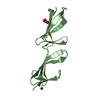
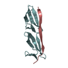
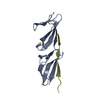
[English] 日本語
Yorodumi
- PDB-2rky: Crystal structure of the fourth and fifth fibronectin F1 modules ... -

+
Open data


ID or keywords:

Loading...

-
Basic information

Entry
Database: PDB / ID: 2rky
TitleCrystal structure of the fourth and fifth fibronectin F1 modules in complex with a fragment of staphylococcus aureus fnbpa-1
Components
  • Fibronectin-binding protein
  • Fibronectin
KeywordsCELL ADHESION / Fibronectin / 4F15F1 / Beta zipper / Staphylococcus aureus / Acute phase / Alternative splicing / Extracellular matrix / Glycoprotein / Heparin-binding / Phosphorylation / Pyrrolidone carboxylic acid / Secreted / Sulfation / Cell wall / Peptidoglycan-anchor / Virulence
Function / homology
Function and homology information


aggregation of unicellular organisms / negative regulation of monocyte activation / calcium-independent cell-matrix adhesion / negative regulation of transforming growth factor beta production / Fibronectin matrix formation / Extracellular matrix organization / positive regulation of substrate-dependent cell migration, cell attachment to substrate / neural crest cell migration involved in autonomic nervous system development / peptidase activator activity / fibrinogen complex ...aggregation of unicellular organisms / negative regulation of monocyte activation / calcium-independent cell-matrix adhesion / negative regulation of transforming growth factor beta production / Fibronectin matrix formation / Extracellular matrix organization / positive regulation of substrate-dependent cell migration, cell attachment to substrate / neural crest cell migration involved in autonomic nervous system development / peptidase activator activity / fibrinogen complex / peptide cross-linking / fibrinogen binding / integrin activation / ALK mutants bind TKIs / cell-substrate junction assembly / biological process involved in interaction with symbiont / Molecules associated with elastic fibres / proteoglycan binding / extracellular matrix structural constituent / MET activates PTK2 signaling / Syndecan interactions / p130Cas linkage to MAPK signaling for integrins / endodermal cell differentiation / GRB2:SOS provides linkage to MAPK signaling for Integrins / fibronectin binding / Signaling by ALK fusions and activated point mutants / Non-integrin membrane-ECM interactions / basement membrane / endoplasmic reticulum-Golgi intermediate compartment / ECM proteoglycans / positive regulation of axon extension / Integrin cell surface interactions / Nuclear events stimulated by ALK signaling in cancer / collagen binding / Degradation of the extracellular matrix / Integrin signaling / substrate adhesion-dependent cell spreading / extracellular matrix / cell-matrix adhesion / regulation of ERK1 and ERK2 cascade / platelet alpha granule lumen / integrin-mediated signaling pathway / acute-phase response / Cell surface interactions at the vascular wall / Post-translational protein phosphorylation / regulation of protein phosphorylation / Signaling by high-kinase activity BRAF mutants / MAP2K and MAPK activation / wound healing / response to wounding / GPER1 signaling / Signaling by RAF1 mutants / Signaling by moderate kinase activity BRAF mutants / Paradoxical activation of RAF signaling by kinase inactive BRAF / Signaling downstream of RAS mutants / Regulation of Insulin-like Growth Factor (IGF) transport and uptake by Insulin-like Growth Factor Binding Proteins (IGFBPs) / positive regulation of fibroblast proliferation / Signaling by BRAF and RAF1 fusions / integrin binding / Platelet degranulation / heparin binding / nervous system development / heart development / regulation of cell shape / angiogenesis / collagen-containing extracellular matrix / Interleukin-4 and Interleukin-13 signaling / blood microparticle / protease binding / positive regulation of phosphatidylinositol 3-kinase/protein kinase B signal transduction / cell adhesion / apical plasma membrane / endoplasmic reticulum lumen / signaling receptor binding / positive regulation of cell population proliferation / positive regulation of gene expression / extracellular space / extracellular exosome / extracellular region / identical protein binding / plasma membrane
Similarity search - Function
Fibronectin binding repeat / Fibronectin binding repeat / Fibrinogen-binding domain 2 / C-terminus of bacterial fibrinogen-binding adhesin / SDR-like Ig domain / Bacterial Ig domain / Fibrogen-binding domain 1 / Fibronectin type I domain / Fibronectin, type I / Fibronectin type-I domain signature. ...Fibronectin binding repeat / Fibronectin binding repeat / Fibrinogen-binding domain 2 / C-terminus of bacterial fibrinogen-binding adhesin / SDR-like Ig domain / Bacterial Ig domain / Fibrogen-binding domain 1 / Fibronectin type I domain / Fibronectin, type I / Fibronectin type-I domain signature. / Fibronectin type-I domain profile. / Fibronectin type 1 domain / Complement Module, domain 1 / Complement Module; domain 1 / Fibronectin type II domain / Fibronectin type II domain superfamily / Fibronectin type II domain / Fibronectin type-II collagen-binding domain signature. / Fibronectin type-II collagen-binding domain profile. / Fibronectin type 2 domain / Adhesion domain superfamily / YSIRK type signal peptide / YSIRK Gram-positive signal peptide / LPXTG cell wall anchor motif / Gram-positive cocci surface proteins LPxTG motif profile. / LPXTG cell wall anchor domain / Kringle-like fold / EGF-like domain signature 1. / Fibronectin type III domain / Fibronectin type 3 domain / Fibronectin type-III domain profile. / Fibronectin type III / Fibronectin type III superfamily / Ribbon / Immunoglobulin-like fold / Mainly Beta
Similarity search - Domain/homology
: / THIOCYANATE ION / Fibronectin / Fibronectin-binding protein A
Similarity search - Component
Biological speciesHomo sapiens (human)
MethodX-RAY DIFFRACTION / SYNCHROTRON / Resolution: 1.8 Å
AuthorsBingham, R.J.
CitationJournal: Proc.Natl.Acad.Sci.Usa / Year: 2008
Title: Crystal structures of fibronectin-binding sites from Staphylococcus aureus FnBPA in complex with fibronectin domains
Authors: Bingham, R.J. / Meenan, N.A. / Schwarz-Linek, U. / Turkenburg, J.P. / Garman, E.F. / Potts, J.R.
History
DepositionOct 18, 2007Deposition site: RCSB / Processing site: PDBJ
Revision 1.0Aug 5, 2008Provider: repository / Type: Initial release
Revision 1.1Jul 13, 2011Group: Version format compliance
Revision 1.2Oct 25, 2017Group: Refinement description / Category: software
Revision 1.3Apr 3, 2024Group: Data collection / Database references ...Data collection / Database references / Derived calculations / Refinement description
Category: chem_comp_atom / chem_comp_bond ...chem_comp_atom / chem_comp_bond / database_2 / pdbx_initial_refinement_model / struct_site
Item: _database_2.pdbx_DOI / _database_2.pdbx_database_accession ..._database_2.pdbx_DOI / _database_2.pdbx_database_accession / _struct_site.pdbx_auth_asym_id / _struct_site.pdbx_auth_comp_id / _struct_site.pdbx_auth_seq_id

-
Structure visualization

Structure viewerMolecule:
MolmilJmol/JSmol

Downloads & links

-
Assembly

Deposited unit
A: Fibronectin
C: Fibronectin
B: Fibronectin-binding protein
D: Fibronectin-binding protein
hetero molecules


Theoretical massNumber of molelcules
Total (without water)26,91310
Polymers26,5834
Non-polymers3306
Water4,216234
1
A: Fibronectin
D: Fibronectin-binding protein
hetero molecules


Theoretical massNumber of molelcules
Total (without water)13,4084
Polymers13,2922
Non-polymers1162
Water362
TypeNameSymmetry operationNumber
identity operation1_555x,y,z1
Buried area2580 Å2
ΔGint-9.9 kcal/mol
Surface area6620 Å2
MethodPISA
2
C: Fibronectin
B: Fibronectin-binding protein
hetero molecules


Theoretical massNumber of molelcules
Total (without water)13,5056
Polymers13,2922
Non-polymers2134
Water362
TypeNameSymmetry operationNumber
identity operation1_555x,y,z1
Buried area2610 Å2
ΔGint-9.2 kcal/mol
Surface area6950 Å2
MethodPISA
Unit cell
Length a, b, c (Å)88.596, 36.244, 73.551
Angle α, β, γ (deg.)90.000, 90.000, 90.000
Int Tables number18
Space group name H-MP21212
Components on special symmetry positions
IDModelComponents
11A-251-

HOH

21A-255-

HOH

-
Components

#1: Protein Fibronectin / / Human fibronectin module pair 4F1-5F1


Mass: 10534.699 Da / Num. of mol.: 2 / Fragment: UNP residues 183-275
Source method: isolated from a genetically manipulated source
Details: human / Source: (gene. exp.) Homo sapiens (human) / Gene: FN1 / Production host: 
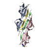
Pichia pastoris (fungus) / Strain (production host): GS115 / References: UniProt: P02751
#2: Protein/peptide Fibronectin-binding protein / Staphylococcus aureus fibronectin binding protein / FNBP


Mass: 2757.012 Da / Num. of mol.: 2 / Fragment: UNP residues 508-530 / Source method: obtained synthetically / Details: Peptide synthesis / References: UniProt: P14738
#3: Chemical
ChemComp-SCN / THIOCYANATE ION / Thiocyanate


Mass: 58.082 Da / Num. of mol.: 5 / Source method: obtained synthetically / Formula: CNS
#4: Chemical ChemComp-K / POTASSIUM ION


Mass: 39.098 Da / Num. of mol.: 1 / Source method: obtained synthetically / Formula: K
#5: Water ChemComp-HOH / water / Water


Mass: 18.015 Da / Num. of mol.: 234 / Source method: isolated from a natural source / Formula: H2O

-
Experimental details

-
Experiment

ExperimentMethod: X-RAY DIFFRACTION / Number of used crystals: 1

-
Sample preparation

CrystalDensity Matthews: 2.22 Å3/Da / Density % sol: 44.62 %
Crystal growTemperature: 291 K / Method: vapor diffusion, hanging drop / pH: 8.5
Details: 100mM Tris-HCl, pH8.5, 0.15M Potassium thiocyanate, 20% PEG400, VAPOR DIFFUSION, HANGING DROP, temperature 291K

-
Data collection

DiffractionMean temperature: 100 K
Diffraction sourceSource: SYNCHROTRON / Site: ESRF / Beamline: ID14-1 / Wavelength: 0.934 Å
DetectorType: ADSC QUANTUM 210 / Detector: CCD / Date: May 9, 2007
RadiationMonochromator: Daimond (111), Ge (220) / Protocol: SINGLE WAVELENGTH / Monochromatic (M) / Laue (L): M / Scattering type: x-ray
Radiation wavelengthWavelength: 0.934 Å / Relative weight: 1
ReflectionResolution: 1.8→30.51 Å / Num. all: 21474 / Num. obs: 21474 / % possible obs: 99.7 % / Observed criterion σ(F): 0 / Observed criterion σ(I): 3.7 / Redundancy: 7.1 % / Biso Wilson estimate: 20.1 Å2 / Rmerge(I) obs: 0.064 / Rsym value: 0.064 / Χ2: 1.858 / Net I/σ(I): 29
Reflection shellResolution: 1.8→1.86 Å / Redundancy: 7.2 % / Rmerge(I) obs: 0.488 / Mean I/σ(I) obs: 6.3 / Num. unique all: 1630 / Rsym value: 0.488 / Χ2: 2.048 / % possible all: 98.97

-
Processing

Software
NameVersionClassificationNB
DENZOdata reduction
SCALEPACKdata scaling
REFMACrefinement
PDB_EXTRACT3data extraction
RefinementStarting model: Previous lower resolution structure

Resolution: 1.8→30.51 Å / Cor.coef. Fo:Fc: 0.954 / Cor.coef. Fo:Fc free: 0.938 / SU B: 2.435 / SU ML: 0.078 / Isotropic thermal model: ISOTROPIC / Cross valid method: THROUGHOUT / σ(F): 0 / σ(I): 3.7 / ESU R: 0.129 / ESU R Free: 0.122 / Stereochemistry target values: MAXIMUM LIKELIHOOD / Details: HYDROGENS HAVE BEEN ADDED IN THE RIDING POSITIONS
RfactorNum. reflection% reflectionSelection details
Rfree0.216 1142 5 %RANDOM
Rwork0.181 ---
all0.183 22616 --
obs0.183 21474 99.7 %-
Solvent computationIon probe radii: 0.8 Å / Shrinkage radii: 0.8 Å / VDW probe radii: 1.2 Å / Solvent model: MASK
Displacement parametersBiso mean: 22.819 Å2
Baniso -1Baniso -2Baniso -3
1-0 Å20 Å20 Å2
2--0.03 Å20 Å2
3----0.03 Å2
Refinement stepCycle: LAST / Resolution: 1.8→30.51 Å
ProteinNucleic acidLigandSolventTotal
Num. atoms1775 0 16 234 2025
Refine LS restraints
Refine-IDTypeDev idealDev ideal targetNumber
X-RAY DIFFRACTIONr_bond_refined_d0.0140.0211827
X-RAY DIFFRACTIONr_angle_refined_deg1.381.922454
X-RAY DIFFRACTIONr_dihedral_angle_1_deg6.2365224
X-RAY DIFFRACTIONr_dihedral_angle_2_deg35.50724.14994
X-RAY DIFFRACTIONr_dihedral_angle_3_deg12.64615325
X-RAY DIFFRACTIONr_dihedral_angle_4_deg12.5021516
X-RAY DIFFRACTIONr_chiral_restr0.0050.02246
X-RAY DIFFRACTIONr_gen_planes_refined0.0060.021394
X-RAY DIFFRACTIONr_nbd_refined0.2290.2278
X-RAY DIFFRACTIONr_nbtor_refined0.2980.2483
X-RAY DIFFRACTIONr_xyhbond_nbd_refined0.1410.285
X-RAY DIFFRACTIONr_symmetry_vdw_refined0.3110.224
X-RAY DIFFRACTIONr_symmetry_hbond_refined0.1840.28
X-RAY DIFFRACTIONr_mcbond_it2.42621108
X-RAY DIFFRACTIONr_mcangle_it3.67331763
X-RAY DIFFRACTIONr_scbond_it3.1532719
X-RAY DIFFRACTIONr_scangle_it4.5243689
LS refinement shellResolution: 1.8→1.847 Å / Total num. of bins used: 20
RfactorNum. reflection% reflection
Rfree0.29 86 -
Rwork0.22 1544 -
all-1630 -
obs-1630 98.97 %

+
About Yorodumi

-
News

-
Feb 9, 2022. New format data for meta-information of EMDB entries

New format data for meta-information of EMDB entries

  • Version 3 of the EMDB header file is now the official format.
  • The previous official version 1.9 will be removed from the archive.

Related info.:EMDB header

External links:wwPDB to switch to version 3 of the EMDB data model

-
Aug 12, 2020. Covid-19 info

Covid-19 info

URL: https://pdbj.org/emnavi/covid19.php

New page: Covid-19 featured information page in EM Navigator.

Related info.:Covid-19 info / Mar 5, 2020. Novel coronavirus structure data

+
Mar 5, 2020. Novel coronavirus structure data

Novel coronavirus structure data

Related info.:Yorodumi Speices / Aug 12, 2020. Covid-19 info

External links:COVID-19 featured content - PDBj / Molecule of the Month (242):Coronavirus Proteases

+
Jan 31, 2019. EMDB accession codes are about to change! (news from PDBe EMDB page)

EMDB accession codes are about to change! (news from PDBe EMDB page)

  • The allocation of 4 digits for EMDB accession codes will soon come to an end. Whilst these codes will remain in use, new EMDB accession codes will include an additional digit and will expand incrementally as the available range of codes is exhausted. The current 4-digit format prefixed with “EMD-” (i.e. EMD-XXXX) will advance to a 5-digit format (i.e. EMD-XXXXX), and so on. It is currently estimated that the 4-digit codes will be depleted around Spring 2019, at which point the 5-digit format will come into force.
  • The EM Navigator/Yorodumi systems omit the EMD- prefix.

Related info.:Q: What is EMD? / ID/Accession-code notation in Yorodumi/EM Navigator

External links:EMDB Accession Codes are Changing Soon! / Contact to PDBj

+
Jul 12, 2017. Major update of PDB

Major update of PDB

  • wwPDB released updated PDB data conforming to the new PDBx/mmCIF dictionary.
  • This is a major update changing the version number from 4 to 5, and with Remediation, in which all the entries are updated.
  • In this update, many items about electron microscopy experimental information are reorganized (e.g. em_software).
  • Now, EM Navigator and Yorodumi are based on the updated data.

External links:wwPDB Remediation / Enriched Model Files Conforming to OneDep Data Standards Now Available in the PDB FTP Archive

-
Yorodumi

Thousand views of thousand structures

  • Yorodumi is a browser for structure data from EMDB, PDB, SASBDB, etc.
  • This page is also the successor to EM Navigator detail page, and also detail information page/front-end page for Omokage search.
  • The word "yorodu" (or yorozu) is an old Japanese word meaning "ten thousand". "mi" (miru) is to see.

Related info.:EMDB / PDB / SASBDB / Comparison of 3 databanks / Yorodumi Search / Aug 31, 2016. New EM Navigator & Yorodumi / Yorodumi Papers / Jmol/JSmol / Function and homology information / Changes in new EM Navigator and Yorodumi

Read more