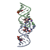
 Movie
Movie Controller


+ Open data

- Basic information
| Entry | Database: PDB / ID: 2wuk | ||||||
|---|---|---|---|---|---|---|---|
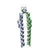
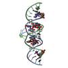
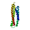
| Title | DivIVA N-terminal domain, F17A mutant | ||||||
 Components Components | SEPTUM SITE-DETERMINING PROTEIN DIVIVA | ||||||
 Keywords Keywords | CELL CYCLE / BACTERIAL CELL DIVISION / SEPTATION / SPORULATION | ||||||
| Function / homology |  Function and homology information Function and homology informationpeptidoglycan-based cell wall biogenesis / division septum assembly / sporulation resulting in formation of a cellular spore / identical protein binding / cytoplasm Similarity search - Function | ||||||
| Biological species |  | ||||||
| Method |  X-RAY DIFFRACTION / MOLECULAR REPLACEMENT / Resolution: 1.9 Å X-RAY DIFFRACTION / MOLECULAR REPLACEMENT / Resolution: 1.9 Å | ||||||
 Authors Authors | Oliva, M.A. / Leonard, T.A. / Lowe, J. | ||||||
 Citation Citation | Journal: Embo J. / Year: 2010 Title: Features Critical for Membrane Binding Revealed by Diviva Crystal Structure. Authors: Oliva, M.A. / Halbedel, S. / Freund, S.M. / Dutow, P. / Leonard, T.A. / Veprintsev, D.B. / Hamoen, L.W. / Lowe, J. | ||||||
| History |
|
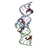
- Structure visualization
| Structure viewer | Molecule: MolmilJmol/JSmol |
|---|
- Downloads & links
-Download
| PDBx/mmCIF format | 2wuk.cif.gz | 64.4 KB | Display | PDBx/mmCIF format |
|---|---|---|---|---|
| PDB format | pdb2wuk.ent.gz | 49.3 KB | Display | PDB format |
| PDBx/mmJSON format | 2wuk.json.gz | Tree view | PDBx/mmJSON format | |
| Others |  Other downloads Other downloads |
-Validation report
| Arichive directory | https://data.pdbj.org/pub/pdb/validation_reports/wu/2wukftp://data.pdbj.org/pub/pdb/validation_reports/wu/2wuk | HTTPS FTP |
|---|
-Related structure data


-Links
 PDBj
PDBj- Assembly
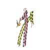
| Deposited unit | 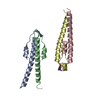
| ||||||||||||
|---|---|---|---|---|---|---|---|---|---|---|---|---|---|
| 1 | 
| ||||||||||||
| 2 | 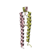
| ||||||||||||
| Unit cell |
| ||||||||||||
| Noncrystallographic symmetry (NCS) | NCS oper:
|
-Components
| #1: Protein | Mass: 6762.587 Da / Num. of mol.: 4 / Fragment: N-TERMINAL DOMAIN, RESIDUES 1-57 / Mutation: YES Source method: isolated from a genetically manipulated source Source: (gene. exp.) #2: Water | ChemComp-HOH / |  Mass: 18.015 Da / Num. of mol.: 388 / Source method: isolated from a natural source / Formula: H2O Mass: 18.015 Da / Num. of mol.: 388 / Source method: isolated from a natural source / Formula: H2OCompound details | ENGINEERED RESIDUE IN CHAIN A, PHE 17 TO ALA ENGINEERED RESIDUE IN CHAIN B, PHE 17 TO ALA ...ENGINEERED | |
|---|
-Experimental details
-Experiment
| Experiment | Method: X-RAY DIFFRACTION / Number of used crystals: 1 |
|---|
- Sample preparation
| Crystal | Density Matthews: 1.9 Å3/Da / Density % sol: 36.2 % / Description: NONE |
|---|
-Data collection
| Diffraction | Mean temperature: 100 K |
|---|---|
| Diffraction source | Source: ROTATING ANODE / Type: RIGAKU RU300 / Wavelength: 1.5418 |
| Detector | Type: MARRESEARCH / Detector: IMAGE PLATE / Date: Feb 1, 2009 |
| Radiation | Protocol: SINGLE WAVELENGTH / Monochromatic (M) / Laue (L): M / Scattering type: x-ray |
| Radiation wavelength | Wavelength: 1.5418 Å / Relative weight: 1 |
| Reflection | Resolution: 1.9→21.3 Å / Num. obs: 14059 / % possible obs: 96.7 % / Observed criterion σ(I): 0 / Redundancy: 2.8 % / Biso Wilson estimate: 16.85 Å2 / Rmerge(I) obs: 0.05 / Net I/σ(I): 13.3 |
| Reflection shell | Resolution: 1.9→2 Å / Redundancy: 2.7 % / Rmerge(I) obs: 0.2 / Mean I/σ(I) obs: 5.2 / % possible all: 96.2 |
- Processing
| Software |
| ||||||||||||||||||||||||||||||||||||||||||||||||||||||||||||||||||||||||||||||||||||
|---|---|---|---|---|---|---|---|---|---|---|---|---|---|---|---|---|---|---|---|---|---|---|---|---|---|---|---|---|---|---|---|---|---|---|---|---|---|---|---|---|---|---|---|---|---|---|---|---|---|---|---|---|---|---|---|---|---|---|---|---|---|---|---|---|---|---|---|---|---|---|---|---|---|---|---|---|---|---|---|---|---|---|---|---|---|
| Refinement | Method to determine structure: MOLECULAR REPLACEMENT / Resolution: 1.9→15.551 Å / SU ML: 0.8 / σ(F): 0.96 / Phase error: 23.45 / Stereochemistry target values: ML
| ||||||||||||||||||||||||||||||||||||||||||||||||||||||||||||||||||||||||||||||||||||
| Solvent computation | Shrinkage radii: 0.9 Å / VDW probe radii: 1.11 Å / Solvent model: FLAT BULK SOLVENT MODEL / Bsol: 60.14 Å2 / ksol: 0.384 e/Å3 | ||||||||||||||||||||||||||||||||||||||||||||||||||||||||||||||||||||||||||||||||||||
| Displacement parameters |
| ||||||||||||||||||||||||||||||||||||||||||||||||||||||||||||||||||||||||||||||||||||
| Refinement step | Cycle: LAST / Resolution: 1.9→15.551 Å
| ||||||||||||||||||||||||||||||||||||||||||||||||||||||||||||||||||||||||||||||||||||
| Refine LS restraints |
| ||||||||||||||||||||||||||||||||||||||||||||||||||||||||||||||||||||||||||||||||||||
| LS refinement shell |
|
