 Movie
Movie Controller


+ Open data

- Basic information
| Entry | Database: PDB / ID: 2wuj | ||||||
|---|---|---|---|---|---|---|---|
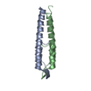
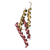
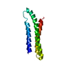
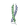
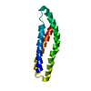
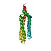
| Title | DivIVA N-terminal domain | ||||||
 Components Components | SEPTUM SITE-DETERMINING PROTEIN DIVIVA | ||||||
 Keywords Keywords | CELL CYCLE / BACTERIAL CELL DIVISION / SEPTATION / SPORULATION | ||||||
| Function / homology |  Function and homology information Function and homology informationpeptidoglycan-based cell wall biogenesis / division septum assembly / sporulation resulting in formation of a cellular spore / identical protein binding / cytoplasm Similarity search - Function | ||||||
| Biological species |  | ||||||
| Method |  X-RAY DIFFRACTION / SYNCHROTRON / SAD / Resolution: 1.4 Å X-RAY DIFFRACTION / SYNCHROTRON / SAD / Resolution: 1.4 Å | ||||||
 Authors Authors | Oliva, M.A. / Leonard, T.A. / Lowe, J. | ||||||
 Citation Citation | Journal: Embo J. / Year: 2010 Title: Features Critical for Membrane Binding Revealed by Diviva Crystal Structure. Authors: Oliva, M.A. / Halbedel, S. / Freund, S.M. / Dutow, P. / Leonard, T.A. / Veprintsev, D.B. / Hamoen, L.W. / Lowe, J. | ||||||
| History |
|
- Structure visualization
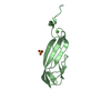
| Structure viewer | Molecule: MolmilJmol/JSmol |
|---|
- Downloads & links
-Download
| PDBx/mmCIF format | 2wuj.cif.gz | 37.6 KB | Display | PDBx/mmCIF format |
|---|---|---|---|---|
| PDB format | pdb2wuj.ent.gz | 26.4 KB | Display | PDB format |
| PDBx/mmJSON format | 2wuj.json.gz | Tree view | PDBx/mmJSON format | |
| Others |  Other downloads Other downloads |
-Validation report
| Arichive directory | https://data.pdbj.org/pub/pdb/validation_reports/wu/2wujftp://data.pdbj.org/pub/pdb/validation_reports/wu/2wuj | HTTPS FTP |
|---|
-Related structure data


-Links
 PDBj
PDBj- Assembly
| Deposited unit | 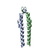
| ||||||||
|---|---|---|---|---|---|---|---|---|---|
| 1 |
| ||||||||
| Unit cell |
| ||||||||
| Noncrystallographic symmetry (NCS) | NCS oper: (Code: given / Matrix: (1), |
-Components
| #1: Protein | Mass: 6838.684 Da / Num. of mol.: 2 / Fragment: N-TERMINAL DOMAIN, RESIDUES 1-57 Source method: isolated from a genetically manipulated source Source: (gene. exp.) #2: Water | ChemComp-HOH / |  Mass: 18.015 Da / Num. of mol.: 211 / Source method: isolated from a natural source / Formula: H2O Mass: 18.015 Da / Num. of mol.: 211 / Source method: isolated from a natural source / Formula: H2O |
|---|
-Experimental details
-Experiment
| Experiment | Method: X-RAY DIFFRACTION / Number of used crystals: 1 |
|---|
- Sample preparation
| Crystal | Density Matthews: 2.3 Å3/Da / Density % sol: 45.4 % / Description: NONE |
|---|
-Data collection
| Diffraction | Mean temperature: 100 K |
|---|---|
| Diffraction source | Source: SYNCHROTRON / Site: ESRF  / Beamline: ID29 / Wavelength: 0.984 / Beamline: ID29 / Wavelength: 0.984 |
| Detector | Type: ADSC CCD / Detector: CCD / Date: Oct 1, 2008 |
| Radiation | Protocol: SINGLE WAVELENGTH / Monochromatic (M) / Laue (L): M / Scattering type: x-ray |
| Radiation wavelength | Wavelength: 0.984 Å / Relative weight: 1 |
| Reflection | Resolution: 1.4→33 Å / Num. obs: 22664 / % possible obs: 94.5 % / Observed criterion σ(I): 0 / Redundancy: 3.3 % / Biso Wilson estimate: 12.7 Å2 / Rmerge(I) obs: 0.07 / Net I/σ(I): 11.2 |
| Reflection shell | Resolution: 1.4→1.48 Å / Redundancy: 2.3 % / Rmerge(I) obs: 0.14 / Mean I/σ(I) obs: 4.8 / % possible all: 88.7 |
- Processing
| Software |
| ||||||||||||||||||||||||||||||||||||||||||||||||||||||||||||||||||||||||||||||||||||||||||||||||||||||||||||||||||||||||||||||||||||||||||||||||||||||||||||||||||||||||||||||||||||||
|---|---|---|---|---|---|---|---|---|---|---|---|---|---|---|---|---|---|---|---|---|---|---|---|---|---|---|---|---|---|---|---|---|---|---|---|---|---|---|---|---|---|---|---|---|---|---|---|---|---|---|---|---|---|---|---|---|---|---|---|---|---|---|---|---|---|---|---|---|---|---|---|---|---|---|---|---|---|---|---|---|---|---|---|---|---|---|---|---|---|---|---|---|---|---|---|---|---|---|---|---|---|---|---|---|---|---|---|---|---|---|---|---|---|---|---|---|---|---|---|---|---|---|---|---|---|---|---|---|---|---|---|---|---|---|---|---|---|---|---|---|---|---|---|---|---|---|---|---|---|---|---|---|---|---|---|---|---|---|---|---|---|---|---|---|---|---|---|---|---|---|---|---|---|---|---|---|---|---|---|---|---|---|---|
| Refinement | Method to determine structure: SAD Starting model: NONE Resolution: 1.4→67.2 Å / Cor.coef. Fo:Fc: 0.949 / Cor.coef. Fo:Fc free: 0.933 / SU B: 0.913 / SU ML: 0.037 / Cross valid method: THROUGHOUT / ESU R: 0.063 / ESU R Free: 0.068 / Stereochemistry target values: MAXIMUM LIKELIHOOD Details: HYDROGENS HAVE BEEN ADDED IN THE RIDING POSITIONS. U VALUES REFINED INDIVIDUALLY.
| ||||||||||||||||||||||||||||||||||||||||||||||||||||||||||||||||||||||||||||||||||||||||||||||||||||||||||||||||||||||||||||||||||||||||||||||||||||||||||||||||||||||||||||||||||||||
| Solvent computation | Ion probe radii: 0.8 Å / Shrinkage radii: 0.8 Å / VDW probe radii: 1.4 Å / Solvent model: BABINET MODEL WITH MASK | ||||||||||||||||||||||||||||||||||||||||||||||||||||||||||||||||||||||||||||||||||||||||||||||||||||||||||||||||||||||||||||||||||||||||||||||||||||||||||||||||||||||||||||||||||||||
| Displacement parameters | Biso mean: 23.766 Å2
| ||||||||||||||||||||||||||||||||||||||||||||||||||||||||||||||||||||||||||||||||||||||||||||||||||||||||||||||||||||||||||||||||||||||||||||||||||||||||||||||||||||||||||||||||||||||
| Refinement step | Cycle: LAST / Resolution: 1.4→67.2 Å
| ||||||||||||||||||||||||||||||||||||||||||||||||||||||||||||||||||||||||||||||||||||||||||||||||||||||||||||||||||||||||||||||||||||||||||||||||||||||||||||||||||||||||||||||||||||||
| Refine LS restraints |
|