 Movie
Movie Controller

[English] 日本語
 Yorodumi
- PDB-7n8i: SARS-CoV-2 S (B.1.429 / epsilon variant) + S2M11 + S2L20 (Local R... -
+ Open data

- Basic information
| Entry | Database: PDB / ID: 7n8i | ||||||
|---|---|---|---|---|---|---|---|
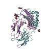
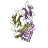
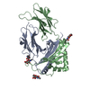
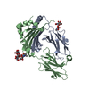
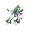
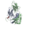
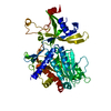
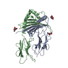
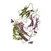
| Title | SARS-CoV-2 S (B.1.429 / epsilon variant) + S2M11 + S2L20 (Local Refinement of the NTD/S2L20) | ||||||
 Components Components |
| ||||||
 Keywords Keywords | VIRAL PROTEIN/IMMUNE SYSTEM / coronavirus / antibody / california / Structural Genomics / Seattle Structural Genomics Center for Infectious Disease / SSGCID / VIRAL PROTEIN / VIRAL PROTEIN-IMMUNE SYSTEM complex | ||||||
| Function / homology |  Function and homology information Function and homology informationsymbiont-mediated disruption of host tissue / Maturation of spike protein / Translation of Structural Proteins / Virion Assembly and Release / host cell surface / host extracellular space / viral translation / symbiont-mediated-mediated suppression of host tetherin activity / Induction of Cell-Cell Fusion / structural constituent of virion ...symbiont-mediated disruption of host tissue / Maturation of spike protein / Translation of Structural Proteins / Virion Assembly and Release / host cell surface / host extracellular space / viral translation / symbiont-mediated-mediated suppression of host tetherin activity / Induction of Cell-Cell Fusion / structural constituent of virion / entry receptor-mediated virion attachment to host cell / membrane fusion / Attachment and Entry / host cell endoplasmic reticulum-Golgi intermediate compartment membrane / positive regulation of viral entry into host cell / receptor-mediated virion attachment to host cell / host cell surface receptor binding / symbiont-mediated suppression of host innate immune response / receptor ligand activity / endocytosis involved in viral entry into host cell / fusion of virus membrane with host plasma membrane / fusion of virus membrane with host endosome membrane / viral envelope / symbiont entry into host cell / virion attachment to host cell / SARS-CoV-2 activates/modulates innate and adaptive immune responses / host cell plasma membrane / virion membrane / identical protein binding / membrane / plasma membrane Similarity search - Function | ||||||
| Biological species |  Homo sapiens (human) Homo sapiens (human)  Severe acute respiratory syndrome coronavirus 2 Severe acute respiratory syndrome coronavirus 2 | ||||||
| Method | ELECTRON MICROSCOPY / single particle reconstruction / cryo EM / Resolution: 3 Å | ||||||
 Authors Authors | McCallum, M. / Seattle Structural Genomics Center for Infectious Disease (SSGCID) / Veesler, D. | ||||||
| Funding support |  United States, 1items United States, 1items
| ||||||
 Citation Citation | Journal: Science / Year: 2021 Title: SARS-CoV-2 immune evasion by the B.1.427/B.1.429 variant of concern. Authors: Matthew McCallum / Jessica Bassi / Anna De Marco / Alex Chen / Alexandra C Walls / Julia Di Iulio / M Alejandra Tortorici / Mary-Jane Navarro / Chiara Silacci-Fregni / Christian Saliba / ...Authors: Matthew McCallum / Jessica Bassi / Anna De Marco / Alex Chen / Alexandra C Walls / Julia Di Iulio / M Alejandra Tortorici / Mary-Jane Navarro / Chiara Silacci-Fregni / Christian Saliba / Kaitlin R Sprouse / Maria Agostini / Dora Pinto / Katja Culap / Siro Bianchi / Stefano Jaconi / Elisabetta Cameroni / John E Bowen / Sasha W Tilles / Matteo Samuele Pizzuto / Sonja Bernasconi Guastalla / Giovanni Bona / Alessandra Franzetti Pellanda / Christian Garzoni / Wesley C Van Voorhis / Laura E Rosen / Gyorgy Snell / Amalio Telenti / Herbert W Virgin / Luca Piccoli / Davide Corti / David Veesler /  Abstract: A novel variant of concern (VOC) named CAL.20C (B.1.427/B.1.429), which was originally detected in California, carries spike glycoprotein mutations S13I in the signal peptide, W152C in the N-terminal ...A novel variant of concern (VOC) named CAL.20C (B.1.427/B.1.429), which was originally detected in California, carries spike glycoprotein mutations S13I in the signal peptide, W152C in the N-terminal domain (NTD), and L452R in the receptor-binding domain (RBD). Plasma from individuals vaccinated with a Wuhan-1 isolate-based messenger RNA vaccine or from convalescent individuals exhibited neutralizing titers that were reduced 2- to 3.5-fold against the B.1.427/B.1.429 variant relative to wild-type pseudoviruses. The L452R mutation reduced neutralizing activity in 14 of 34 RBD-specific monoclonal antibodies (mAbs). The S13I and W152C mutations resulted in total loss of neutralization for 10 of 10 NTD-specific mAbs because the NTD antigenic supersite was remodeled by a shift of the signal peptide cleavage site and the formation of a new disulfide bond, as revealed by mass spectrometry and structural studies. | ||||||
| History |
|
- Structure visualization
| Movie |
Movie viewer |
|---|---|
| Structure viewer | Molecule: MolmilJmol/JSmol |
- Downloads & links
-Download
| PDBx/mmCIF format | 7n8i.cif.gz | 122.4 KB | Display | PDBx/mmCIF format |
|---|---|---|---|---|
| PDB format | pdb7n8i.ent.gz | 80 KB | Display | PDB format |
| PDBx/mmJSON format | 7n8i.json.gz | Tree view | PDBx/mmJSON format | |
| Others |  Other downloads Other downloads |
-Validation report
| Summary document | 7n8i_validation.pdf.gz | 1.1 MB | Display | wwPDB validaton report |
|---|---|---|---|---|
| Full document | 7n8i_full_validation.pdf.gz | 1.1 MB | Display | |
| Data in XML | 7n8i_validation.xml.gz | 31.5 KB | Display | |
| Data in CIF | 7n8i_validation.cif.gz | 42.3 KB | Display | |
| Arichive directory | https://data.pdbj.org/pub/pdb/validation_reports/n8/7n8iftp://data.pdbj.org/pub/pdb/validation_reports/n8/7n8i | HTTPS FTP |
-Related structure data
| Related structure data |  24237MC  7n8hC C: citing same article ( M: map data used to model this data |
|---|---|
| Similar structure data |


-Links
 PDBj
PDBj


- Assembly
| Deposited unit | 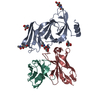
|
|---|---|
| 1 |
|
-Components
| #1: Antibody | Mass: 11699.961 Da / Num. of mol.: 1 Source method: isolated from a genetically manipulated source Source: (gene. exp.) Homo sapiens (human) / Production host: Homo sapiens (human) | ||||
|---|---|---|---|---|---|
| #2: Antibody | Mass: 13453.958 Da / Num. of mol.: 1 Source method: isolated from a genetically manipulated source Source: (gene. exp.) Homo sapiens (human) / Production host: Homo sapiens (human) | ||||
| #3: Protein | Mass: 141484.609 Da / Num. of mol.: 1 Source method: isolated from a genetically manipulated source Source: (gene. exp.) Severe acute respiratory syndrome coronavirus 2Gene: S, 2 / Production host: Homo sapiens (human) / References: UniProt: P0DTC2 | ||||
| #4: Sugar | ChemComp-NAG /   Type: D-saccharide, beta linking / Mass: 221.208 Da / Num. of mol.: 5 / Source method: obtained synthetically / Formula: C8H15NO6 Type: D-saccharide, beta linking / Mass: 221.208 Da / Num. of mol.: 5 / Source method: obtained synthetically / Formula: C8H15NO6Has ligand of interest | N | Has protein modification | Y | |
-Experimental details
-Experiment
| Experiment | Method: ELECTRON MICROSCOPY |
|---|---|
| EM experiment | Aggregation state: PARTICLE / 3D reconstruction method: single particle reconstruction |
- Sample preparation
| Component | Name: SARS-CoV-2 S (B.1.429 / epsilon variant) bound to S2L20 Fab Type: COMPLEX / Entity ID: #1-#3 / Source: RECOMBINANT | ||||||||||||
|---|---|---|---|---|---|---|---|---|---|---|---|---|---|
| Source (natural) |
| ||||||||||||
| Source (recombinant) | Organism: Homo sapiens (human) | ||||||||||||
| Buffer solution | pH: 8.5 | ||||||||||||
| Specimen | Embedding applied: NO / Shadowing applied: NO / Staining applied: NO / Vitrification applied: YES | ||||||||||||
| Vitrification | Cryogen name: ETHANE |
- Electron microscopy imaging
| Experimental equipment |  Model: Titan Krios / Image courtesy: FEI Company |
|---|---|
| Microscopy | Model: FEI TITAN KRIOS |
| Electron gun | Electron source:  FIELD EMISSION GUN / Accelerating voltage: 300 kV / Illumination mode: FLOOD BEAM FIELD EMISSION GUN / Accelerating voltage: 300 kV / Illumination mode: FLOOD BEAM |
| Electron lens | Mode: BRIGHT FIELD |
| Image recording | Electron dose: 63 e/Å2 / Film or detector model: GATAN K3 (6k x 4k) |
- Processing
| CTF correction | Type: PHASE FLIPPING AND AMPLITUDE CORRECTION |
|---|---|
| 3D reconstruction | Resolution: 3 Å / Resolution method: FSC 0.143 CUT-OFF / Num. of particles: 254593 / Symmetry type: POINT |
