 Movie
Movie Controller


+ Open data

- Basic information
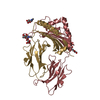
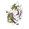
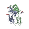
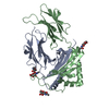
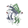
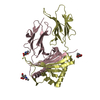
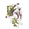
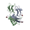
| Entry | Database: PDB / ID: 1ieb | |||||||||
|---|---|---|---|---|---|---|---|---|---|---|
| Title | HISTOCOMPATIBILITY ANTIGEN | |||||||||
 Components Components | (MHC CLASS II I-EK) x 2 | |||||||||
 Keywords Keywords | HISTOCOMPATIBILITY ANTIGEN | |||||||||
| Function / homology |  Function and homology information Function and homology informationPhosphorylation of CD3 and TCR zeta chains / Translocation of ZAP-70 to Immunological synapse / Co-inhibition by PD-1 / Generation of second messenger molecules / Downstream TCR signaling / myeloid dendritic cell antigen processing and presentation / antigen processing and presentation of endogenous peptide antigen via MHC class II / regulation of T-helper cell differentiation / positive regulation of CD4-positive, CD25-positive, alpha-beta regulatory T cell differentiation / positive regulation of CD4-positive, alpha-beta T cell activation ...Phosphorylation of CD3 and TCR zeta chains / Translocation of ZAP-70 to Immunological synapse / Co-inhibition by PD-1 / Generation of second messenger molecules / Downstream TCR signaling / myeloid dendritic cell antigen processing and presentation / antigen processing and presentation of endogenous peptide antigen via MHC class II / regulation of T-helper cell differentiation / positive regulation of CD4-positive, CD25-positive, alpha-beta regulatory T cell differentiation / positive regulation of CD4-positive, alpha-beta T cell activation / antigen processing and presentation of peptide or polysaccharide antigen via MHC class II / positive regulation of memory T cell differentiation / MHC class II antigen presentation / immunoglobulin mediated immune response / polysaccharide binding / immunological synapse / T cell receptor binding / peptide antigen assembly with MHC class II protein complex / MHC class II protein complex / positive regulation of T cell mediated cytotoxicity / antigen processing and presentation of exogenous peptide antigen via MHC class II / positive regulation of immune response / peptide antigen binding / cognition / positive regulation of T cell activation / MHC class II protein complex binding / late endosome membrane / adaptive immune response / lysosome / external side of plasma membrane / lysosomal membrane / cell surface / plasma membrane Similarity search - Function | |||||||||
| Biological species |  | |||||||||
| Method |  X-RAY DIFFRACTION / SYNCHROTRON / MOLECULAR REPLACEMENT / Resolution: 2.7 Å X-RAY DIFFRACTION / SYNCHROTRON / MOLECULAR REPLACEMENT / Resolution: 2.7 Å | |||||||||
 Authors Authors | Fremont, D.H. / Hendrickson, W.A. / Marrack, P. / Kappler, J. | |||||||||
 Citation Citation | Journal: Science / Year: 1996 Title: Structures of an MHC class II molecule with covalently bound single peptides. Authors: Fremont, D.H. / Hendrickson, W.A. / Marrack, P. / Kappler, J. | |||||||||
| History |
|
- Structure visualization
| Structure viewer | Molecule: MolmilJmol/JSmol |
|---|
- Downloads & links
-Download
| PDBx/mmCIF format | 1ieb.cif.gz | 176.7 KB | Display | PDBx/mmCIF format |
|---|---|---|---|---|
| PDB format | pdb1ieb.ent.gz | 140.4 KB | Display | PDB format |
| PDBx/mmJSON format | 1ieb.json.gz | Tree view | PDBx/mmJSON format | |
| Others |  Other downloads Other downloads |
-Validation report
| Arichive directory | https://data.pdbj.org/pub/pdb/validation_reports/ie/1iebftp://data.pdbj.org/pub/pdb/validation_reports/ie/1ieb | HTTPS FTP |
|---|
-Related structure data
| Related structure data |  1ieaSC S: Starting model for refinement C: citing same article ( |
|---|---|
| Similar structure data |
-Links
 PDBj
PDBj


- Assembly
| Deposited unit | 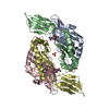
| ||||||||
|---|---|---|---|---|---|---|---|---|---|
| 1 | 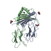
| ||||||||
| 2 | 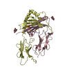
| ||||||||
| 3 |
| ||||||||
| Unit cell |
| ||||||||
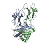
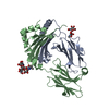
| Details | THERE ARE TWO HETERODIMERS IN THE ASYMMETRIC UNIT RELATED BY AN APPROXIMATE TWO-FOLD AXIS. MOLECULE 1 : ALPHA CHAIN - RESIDUES A 1 THROUGH A 182 : BETA CHAIN - RESIDUES B 4N THROUGH B 188 : SUGARS - RESIDUES A 1, A 2, B 3 MOLECULE 2 : ALPHA CHAIN - RESIDUES C 1 THROUGH A 182 : BETA CHAIN - RESIDUES D 4N THROUGH B 188 : SUGARS - RESIDUES C 4, C 5, D 6 WATERS : 1 THROUGH 159 |
-Components
| #1: Protein | Mass: 22324.008 Da / Num. of mol.: 2 Source method: isolated from a genetically manipulated source Details: WITH COVALENTLY BOUND HSP70 PEPTIDE / Source: (gene. exp.)   Spodoptera frugiperda (fall armyworm) / References: UniProt: P01904 Spodoptera frugiperda (fall armyworm) / References: UniProt: P01904#2: Protein | Mass: 26307.381 Da / Num. of mol.: 2 Source method: isolated from a genetically manipulated source Details: WITH COVALENTLY BOUND HSP70 PEPTIDE / Source: (gene. exp.) Spodoptera frugiperda (fall armyworm) / References: UniProt: Q31164#3: Sugar | ChemComp-NAG /   Type: D-saccharide, beta linking / Mass: 221.208 Da / Num. of mol.: 6 Type: D-saccharide, beta linking / Mass: 221.208 Da / Num. of mol.: 6Source method: isolated from a genetically manipulated source Formula: C8H15NO6 #4: Chemical | ChemComp-SO4 / |   Mass: 96.063 Da / Num. of mol.: 1 / Source method: obtained synthetically / Formula: SO4 Mass: 96.063 Da / Num. of mol.: 1 / Source method: obtained synthetically / Formula: SO4#5: Water | ChemComp-HOH / |  Mass: 18.015 Da / Num. of mol.: 159 / Source method: isolated from a natural source / Formula: H2O Mass: 18.015 Da / Num. of mol.: 159 / Source method: isolated from a natural source / Formula: H2OCompound details | THIS ENTRY CONTAINS COORDINATES FOR THE EXTRACELLULAR DOMAINS OF THE MURINE MHC CLASS II MOLECULE I- ...THIS ENTRY CONTAINS COORDINATE | Has protein modification | Y | |
|---|
-Experimental details
-Experiment
| Experiment | Method: X-RAY DIFFRACTION / Number of used crystals: 1 |
|---|
- Sample preparation
| Crystal | Density Matthews: 2.85 Å3/Da / Density % sol: 60 % | ||||||||||||||||||||||||||||||
|---|---|---|---|---|---|---|---|---|---|---|---|---|---|---|---|---|---|---|---|---|---|---|---|---|---|---|---|---|---|---|---|
| Crystal grow | pH: 5.2 Details: 18% PEG 4000, 2% EG, 200MM A.S, 100MM CITRATE PH 5.2 | ||||||||||||||||||||||||||||||
| Crystal grow | *PLUS Method: unknown / PH range low: 5.6 / PH range high: 5.2 | ||||||||||||||||||||||||||||||
| Components of the solutions | *PLUS
|
-Data collection
| Diffraction | Mean temperature: 110 K |
|---|---|
| Diffraction source | Source: SYNCHROTRON / Site: NSLS  / Beamline: X4A / Wavelength: 0.9789 / Beamline: X4A / Wavelength: 0.9789 |
| Detector | Type: FUJI / Detector: IMAGE PLATE |
| Radiation | Scattering type: x-ray |
| Radiation wavelength | Wavelength: 0.9789 Å / Relative weight: 1 |
| Reflection | Resolution: 2.7→20 Å / Num. obs: 29069 / % possible obs: 92.1 % / Redundancy: 2.95 % / Rsym value: 0.087 |
| Reflection | *PLUS Num. measured all: 85623 / Rmerge(I) obs: 0.087 |
- Processing
| Software |
| ||||||||||||||||||||||||||||||||||||||||||||||||||||||||||||||||||||||||||||||||
|---|---|---|---|---|---|---|---|---|---|---|---|---|---|---|---|---|---|---|---|---|---|---|---|---|---|---|---|---|---|---|---|---|---|---|---|---|---|---|---|---|---|---|---|---|---|---|---|---|---|---|---|---|---|---|---|---|---|---|---|---|---|---|---|---|---|---|---|---|---|---|---|---|---|---|---|---|---|---|---|---|---|
| Refinement | Method to determine structure: MOLECULAR REPLACEMENT Starting model: PDB ENTRY 1IEA Resolution: 2.7→6 Å / Rfactor Rfree error: 0.009 / Data cutoff high absF: 100000 / Data cutoff low absF: 0.01 / Cross valid method: THROUGHOUT / σ(F): 2
| ||||||||||||||||||||||||||||||||||||||||||||||||||||||||||||||||||||||||||||||||
| Displacement parameters | Biso mean: 36.5 Å2 | ||||||||||||||||||||||||||||||||||||||||||||||||||||||||||||||||||||||||||||||||
| Refine analyze | Luzzati coordinate error obs: 0.34 Å | ||||||||||||||||||||||||||||||||||||||||||||||||||||||||||||||||||||||||||||||||
| Refinement step | Cycle: LAST / Resolution: 2.7→6 Å
| ||||||||||||||||||||||||||||||||||||||||||||||||||||||||||||||||||||||||||||||||
| Refine LS restraints |
| ||||||||||||||||||||||||||||||||||||||||||||||||||||||||||||||||||||||||||||||||
| Refine LS restraints NCS | NCS model details: RESTRAINED | ||||||||||||||||||||||||||||||||||||||||||||||||||||||||||||||||||||||||||||||||
| LS refinement shell | Resolution: 2.7→2.85 Å / Rfactor Rfree error: 0.03 / Total num. of bins used: 6
| ||||||||||||||||||||||||||||||||||||||||||||||||||||||||||||||||||||||||||||||||
| Xplor file |
| ||||||||||||||||||||||||||||||||||||||||||||||||||||||||||||||||||||||||||||||||
| Software | *PLUS Name: X-PLOR / Classification: refinement | ||||||||||||||||||||||||||||||||||||||||||||||||||||||||||||||||||||||||||||||||
| Refine LS restraints | *PLUS
|
