 Movie
Movie Controller


+ Open data

- Basic information
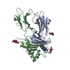
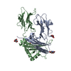
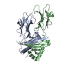
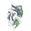
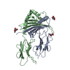
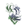
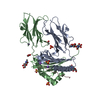
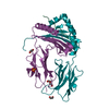
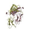
| Entry | Database: PDB / ID: 1fne | ||||||
|---|---|---|---|---|---|---|---|
| Title | HISTOCOMPATIBILITY ANTIGEN | ||||||
 Components Components |
| ||||||
 Keywords Keywords | IMMUNE SYSTEM / HISTOCOMPATIBILITY ANTIGEN / MHC | ||||||
| Function / homology |  Function and homology information Function and homology informationhemoglobin beta binding / positive regulation of myeloid cell differentiation / myeloid dendritic cell antigen processing and presentation / antigen processing and presentation of endogenous peptide antigen via MHC class II / regulation of T-helper cell differentiation / positive regulation of CD4-positive, CD25-positive, alpha-beta regulatory T cell differentiation / positive regulation of CD4-positive, alpha-beta T cell activation / antigen processing and presentation of peptide or polysaccharide antigen via MHC class II / positive regulation of memory T cell differentiation / cellular oxidant detoxification ...hemoglobin beta binding / positive regulation of myeloid cell differentiation / myeloid dendritic cell antigen processing and presentation / antigen processing and presentation of endogenous peptide antigen via MHC class II / regulation of T-helper cell differentiation / positive regulation of CD4-positive, CD25-positive, alpha-beta regulatory T cell differentiation / positive regulation of CD4-positive, alpha-beta T cell activation / antigen processing and presentation of peptide or polysaccharide antigen via MHC class II / positive regulation of memory T cell differentiation / cellular oxidant detoxification / nitric oxide transport / hemoglobin alpha binding / hemoglobin binding / haptoglobin-hemoglobin complex / hemoglobin complex / immunoglobulin mediated immune response / polysaccharide binding / oxygen transport / immunological synapse / erythrocyte development / T cell receptor binding / oxygen carrier activity / glutathione metabolic process / carbon dioxide transport / regulation of erythrocyte differentiation / oxygen binding / peptide antigen assembly with MHC class II protein complex / MHC class II protein complex / positive regulation of T cell mediated cytotoxicity / antigen processing and presentation of exogenous peptide antigen via MHC class II / positive regulation of immune response / peptide antigen binding / cognition / positive regulation of T cell activation / MHC class II protein complex binding / late endosome membrane / lysosome / external side of plasma membrane / lysosomal membrane / heme binding / protein-containing complex binding / cell surface / : / metal ion binding / plasma membrane Similarity search - Function | ||||||
| Biological species |  | ||||||
| Method |  X-RAY DIFFRACTION / SYNCHROTRON / MOLECULAR REPLACEMENT / Resolution: 1.9 Å X-RAY DIFFRACTION / SYNCHROTRON / MOLECULAR REPLACEMENT / Resolution: 1.9 Å | ||||||
 Authors Authors | Miley, M.J. / Nelson, C.A. / Fremont, D.H. | ||||||
 Citation Citation | Journal: J.Immunol. / Year: 2001 Title: Structural and functional consequences of altering a peptide MHC anchor residue. Authors: Kersh, G.J. / Miley, M.J. / Nelson, C.A. / Grakoui, A. / Horvath, S. / Donermeyer, D.L. / Kappler, J. / Allen, P.M. / Fremont, D.H. | ||||||
| History |
|
- Structure visualization
| Structure viewer | Molecule: MolmilJmol/JSmol |
|---|
- Downloads & links
-Download
| PDBx/mmCIF format | 1fne.cif.gz | 183.6 KB | Display | PDBx/mmCIF format |
|---|---|---|---|---|
| PDB format | pdb1fne.ent.gz | 146.4 KB | Display | PDB format |
| PDBx/mmJSON format | 1fne.json.gz | Tree view | PDBx/mmJSON format | |
| Others |  Other downloads Other downloads |
-Validation report
| Arichive directory | https://data.pdbj.org/pub/pdb/validation_reports/fn/1fneftp://data.pdbj.org/pub/pdb/validation_reports/fn/1fne | HTTPS FTP |
|---|
-Related structure data
| Related structure data |  1fngC 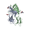 1ieaS S: Starting model for refinement C: citing same article ( |
|---|---|
| Similar structure data |


-Links
 PDBj
PDBj


- Assembly
| Deposited unit | 
| ||||||||
|---|---|---|---|---|---|---|---|---|---|
| 1 | 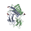
| ||||||||
| 2 | 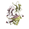
| ||||||||
| 3 | 
| ||||||||
| Unit cell |
|
-Components
| #1: Protein | Mass: 22361.051 Da / Num. of mol.: 2 / Fragment: SOLUBLE ECTO-DOMAIN Source method: isolated from a genetically manipulated source Source: (gene. exp.)   Spodoptera frugiperda (fall armyworm) / References: UniProt: P04224 Spodoptera frugiperda (fall armyworm) / References: UniProt: P04224#2: Protein | Mass: 25362.254 Da / Num. of mol.: 2 Fragment: SOLUBLE ECTO-DOMAIN WITH COVALENTLY ATTACHED HB PEPTIDE Mutation: E(6P)D IN PEPTIDE RESIDUE 6P Source method: isolated from a genetically manipulated source Details: WITH COVALENTLY BOUND HB(D73) PEPTIDE / Source: (gene. exp.) Spodoptera frugiperda (fall armyworm) / References: UniProt: P02089, GenBank: AAA39594#3: Sugar | ChemComp-NAG /   Type: D-saccharide, beta linking / Mass: 221.208 Da / Num. of mol.: 6 / Source method: obtained synthetically / Formula: C8H15NO6 Type: D-saccharide, beta linking / Mass: 221.208 Da / Num. of mol.: 6 / Source method: obtained synthetically / Formula: C8H15NO6#4: Water | ChemComp-HOH / |  Mass: 18.015 Da / Num. of mol.: 507 / Source method: isolated from a natural source / Formula: H2O Mass: 18.015 Da / Num. of mol.: 507 / Source method: isolated from a natural source / Formula: H2OCompound details | THIS ENTRY CONTAINS COORDINATES FOR THE EXTRACELLULAR DOMAINS OF THE MURINE MHC CLASS II MOLECULE I- ...THIS ENTRY CONTAINS COORDINATE | Has protein modification | Y | Sequence details | RESIDUES (1L - 16L)IN CHAINS B & D FORM A POLYPEPTID | |
|---|
-Experimental details
-Experiment
| Experiment | Method: X-RAY DIFFRACTION / Number of used crystals: 3 |
|---|
- Sample preparation
| Crystal | Density Matthews: 2.55 Å3/Da / Density % sol: 51.78 % | ||||||||||||||||||||||||||||||||||||||||
|---|---|---|---|---|---|---|---|---|---|---|---|---|---|---|---|---|---|---|---|---|---|---|---|---|---|---|---|---|---|---|---|---|---|---|---|---|---|---|---|---|---|
| Crystal grow | Temperature: 293 K / Method: vapor diffusion, hanging drop / pH: 4.8 Details: 15% PEG 4000 15% 2-PROPANOL 300mM-500mM AMMONIUM ACETATE 100mM CITRATE PH 4.8, VAPOR DIFFUSION, HANGING DROP, temperature 293K | ||||||||||||||||||||||||||||||||||||||||
| Crystal grow | *PLUS Temperature: 20 ℃ / pH: 7 | ||||||||||||||||||||||||||||||||||||||||
| Components of the solutions | *PLUS
|
-Data collection
| Diffraction | Mean temperature: 110 K |
|---|---|
| Diffraction source | Source: SYNCHROTRON / Site: APS  / Beamline: 19-ID / Wavelength: 1.0332 / Beamline: 19-ID / Wavelength: 1.0332 |
| Detector | Detector: CCD |
| Radiation | Protocol: SINGLE WAVELENGTH / Monochromatic (M) / Laue (L): M / Scattering type: x-ray |
| Radiation wavelength | Wavelength: 1.0332 Å / Relative weight: 1 |
| Reflection | Resolution: 1.9→100 Å / Num. all: 364002 / Num. obs: 72521 / % possible obs: 94.4 % / Redundancy: 5.01 % / Biso Wilson estimate: 29.6 Å2 / Rmerge(I) obs: 0.114 / Rsym value: 0.114 / Net I/σ(I): 13.5 |
| Reflection shell | Resolution: 1.9→1.96 Å / Rmerge(I) obs: 0.301 / % possible all: 92.6 |
| Reflection | *PLUS Num. measured all: 364002 |
| Reflection shell | *PLUS % possible obs: 75.6 % / Mean I/σ(I) obs: 2.45 |
- Processing
| Software |
| ||||||||||||||||||||||||||||||||||||||||
|---|---|---|---|---|---|---|---|---|---|---|---|---|---|---|---|---|---|---|---|---|---|---|---|---|---|---|---|---|---|---|---|---|---|---|---|---|---|---|---|---|---|
| Refinement | Method to determine structure: MOLECULAR REPLACEMENT Starting model: PDB ENTRY 1IEA Resolution: 1.9→19.97 Å / Rfactor Rfree error: 0.004 / Data cutoff high absF: 845534.75 / Data cutoff high rms absF: 845534.75 / Data cutoff low absF: 0 / Isotropic thermal model: RESTRAINED / Cross valid method: THROUGHOUT
| ||||||||||||||||||||||||||||||||||||||||
| Solvent computation | Solvent model: FLAT MODEL / Bsol: 58.25 Å2 / ksol: 0.356 e/Å3 | ||||||||||||||||||||||||||||||||||||||||
| Displacement parameters | Biso mean: 41.4 Å2
| ||||||||||||||||||||||||||||||||||||||||
| Refine analyze |
| ||||||||||||||||||||||||||||||||||||||||
| Refinement step | Cycle: LAST / Resolution: 1.9→19.97 Å
| ||||||||||||||||||||||||||||||||||||||||
| Refine LS restraints |
| ||||||||||||||||||||||||||||||||||||||||
| LS refinement shell | Resolution: 1.9→1.96 Å / Rfactor Rfree error: 0.027 / Total num. of bins used: 12
| ||||||||||||||||||||||||||||||||||||||||
| Xplor file |
| ||||||||||||||||||||||||||||||||||||||||
| Software | *PLUS Name: CNS / Classification: refinement | ||||||||||||||||||||||||||||||||||||||||
| Refinement | *PLUS % reflection Rfree: 5.1 % / Rfactor Rfree: 0.27 | ||||||||||||||||||||||||||||||||||||||||
| Solvent computation | *PLUS | ||||||||||||||||||||||||||||||||||||||||
| Displacement parameters | *PLUS Biso mean: 41.4 Å2 | ||||||||||||||||||||||||||||||||||||||||
| Refine LS restraints | *PLUS
| ||||||||||||||||||||||||||||||||||||||||
| LS refinement shell | *PLUS Rfactor Rfree: 0.42 / % reflection Rfree: 5 % / Rfactor Rwork: 0.36 / Rfactor obs: 0.36 |
