[English] 日本語
Yorodumi
- PDB-7n86: Crystal Structure of Human Protocadherin-24 EC1-2 Form II -

+
Open data


ID or keywords:

Loading...

-
Basic information

Entry
Database: PDB / ID: 7n86
TitleCrystal Structure of Human Protocadherin-24 EC1-2 Form II
ComponentsCadherin-related family member 2
KeywordsCELL ADHESION / BRUSH BORDER / CALCIUM BINDING PROTEIN
Function / homology
Function and homology information


negative regulation of cell growth involved in contact inhibition / intermicrovillar adhesion / regulation of microvillus length / cell-cell adhesion mediated by cadherin / anchoring junction / microvillus membrane / homophilic cell adhesion via plasma membrane adhesion molecules / brush border / cell adhesion molecule binding / epithelial cell differentiation ...negative regulation of cell growth involved in contact inhibition / intermicrovillar adhesion / regulation of microvillus length / cell-cell adhesion mediated by cadherin / anchoring junction / microvillus membrane / homophilic cell adhesion via plasma membrane adhesion molecules / brush border / cell adhesion molecule binding / epithelial cell differentiation / brush border membrane / apical plasma membrane / calcium ion binding / extracellular exosome / plasma membrane
Similarity search - Function
Cadherin / Cadherin conserved site / Cadherin domain signature. / Cadherin repeats. / Cadherin domain / Cadherins domain profile. / Cadherin-like / Cadherin-like superfamily
Similarity search - Domain/homology
IODIDE ION / Cadherin-related family member 2
Similarity search - Component
Biological speciesHomo sapiens (human)
MethodX-RAY DIFFRACTION / SYNCHROTRON / MOLECULAR REPLACEMENT / Resolution: 3.175 Å
AuthorsModak, D. / Gray, M.E. / Sotomayor, M.
Funding support United States, 1items
OrganizationGrant numberCountry
National Institutes of Health/National Institute of Diabetes and Digestive and Kidney Disease (NIH/NIDDK)R01 DK095811 United States
CitationJournal: Plos Biol. / Year: 2021
Title: Heterophilic and homophilic cadherin interactions in intestinal intermicrovillar links are species dependent.
Authors: Gray, M.E. / Johnson, Z.R. / Modak, D. / Tamilselvan, E. / Tyska, M.J. / Sotomayor, M.
History
DepositionJun 13, 2021Deposition site: RCSB / Processing site: RCSB
Revision 1.0Sep 1, 2021Provider: repository / Type: Initial release
Revision 1.1Dec 22, 2021Group: Database references / Category: citation / citation_author
Item: _citation.country / _citation.journal_abbrev ..._citation.country / _citation.journal_abbrev / _citation.journal_id_CSD / _citation.journal_id_ISSN / _citation.journal_volume / _citation.page_first / _citation.page_last / _citation.pdbx_database_id_DOI / _citation.pdbx_database_id_PubMed / _citation.title / _citation.year
Revision 1.2Oct 18, 2023Group: Data collection / Refinement description
Category: chem_comp_atom / chem_comp_bond ...chem_comp_atom / chem_comp_bond / pdbx_initial_refinement_model / struct_ncs_dom_lim
Item: _struct_ncs_dom_lim.beg_auth_comp_id / _struct_ncs_dom_lim.beg_label_asym_id ..._struct_ncs_dom_lim.beg_auth_comp_id / _struct_ncs_dom_lim.beg_label_asym_id / _struct_ncs_dom_lim.beg_label_comp_id / _struct_ncs_dom_lim.beg_label_seq_id / _struct_ncs_dom_lim.end_auth_comp_id / _struct_ncs_dom_lim.end_label_asym_id / _struct_ncs_dom_lim.end_label_comp_id / _struct_ncs_dom_lim.end_label_seq_id

-
Structure visualization

Structure viewerMolecule:
MolmilJmol/JSmol

Downloads & links

-
Assembly

Deposited unit
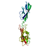
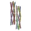
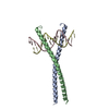
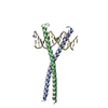
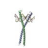
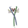
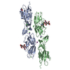
A: Cadherin-related family member 2
B: Cadherin-related family member 2
C: Cadherin-related family member 2
D: Cadherin-related family member 2
hetero molecules


Theoretical massNumber of molelcules
Total (without water)107,58432
Polymers104,1694
Non-polymers3,41528
Water59433
1
A: Cadherin-related family member 2
B: Cadherin-related family member 2
hetero molecules


Theoretical massNumber of molelcules
Total (without water)53,86416
Polymers52,0852
Non-polymers1,77914
Water362
TypeNameSymmetry operationNumber
identity operation1_555x,y,z1
Buried area5210 Å2
ΔGint-38 kcal/mol
Surface area21010 Å2
MethodPISA
2
C: Cadherin-related family member 2
D: Cadherin-related family member 2
hetero molecules


Theoretical massNumber of molelcules
Total (without water)53,72016
Polymers52,0852
Non-polymers1,63614
Water362
TypeNameSymmetry operationNumber
identity operation1_555x,y,z1
Buried area4750 Å2
ΔGint-52 kcal/mol
Surface area21380 Å2
MethodPISA
Unit cell
Length a, b, c (Å)63.899, 86.641, 104.892
Angle α, β, γ (deg.)90.000, 103.458, 90.000
Int Tables number4
Space group name H-MP1211
Noncrystallographic symmetry (NCS)NCS domain:
IDEns-IDDetails
11A
21B
32A
42C
53A
63D
74B
84C
95B
105D
116C
126D

NCS domain segments:

Component-ID: 1 / Beg auth comp-ID: ASN / Beg label comp-ID: ASN / Refine code: 1

Dom-IDEns-IDEnd auth comp-IDEnd label comp-IDAuth asym-IDLabel asym-IDAuth seq-IDLabel seq-ID
11PROPROAA1 - 2151 - 215
21PROPROBB1 - 2151 - 215
32ASPASPAA1 - 2131 - 213
42ASPASPCC1 - 2131 - 213
53GLNGLNAA1 - 2141 - 214
63GLNGLNDD1 - 2141 - 214
74ASPASPBB1 - 2131 - 213
84ASPASPCC1 - 2131 - 213
95GLNGLNBB1 - 2141 - 214
105GLNGLNDD1 - 2141 - 214
116ASPASPCC1 - 2131 - 213
126ASPASPDD1 - 2131 - 213

NCS ensembles :
IDDetails
1Global NCS restraints between domains: 1 2
2Global NCS restraints between domains: 3 4
3Global NCS restraints between domains: 5 6
4Global NCS restraints between domains: 7 8
5Global NCS restraints between domains: 9 10
6Global NCS restraints between domains: 11 12

-
Components

-
Protein / Sugars , 2 types, 8 molecules ABCD

#1: Protein
Cadherin-related family member 2 / Protocadherin LKC / PC-LKC / Protocadherin-24


Mass: 26042.340 Da / Num. of mol.: 4
Source method: isolated from a genetically manipulated source
Details: Cleaved signal peptide (1-20) not included / Source: (gene. exp.) Homo sapiens (human) / Gene: CDHR2, PCDH24, PCLKC / Plasmid: PHIS-N1 / Cell line (production host): EXPI293 / Production host: Homo sapiens (human) / References: UniProt: Q9BYE9
#2: Polysaccharide
beta-D-mannopyranose-(1-4)-2-acetamido-2-deoxy-beta-D-glucopyranose-(1-4)-2-acetamido-2-deoxy-beta- ...beta-D-mannopyranose-(1-4)-2-acetamido-2-deoxy-beta-D-glucopyranose-(1-4)-2-acetamido-2-deoxy-beta-D-glucopyranose


Type: oligosaccharide / Mass: 586.542 Da / Num. of mol.: 4
Source method: isolated from a genetically manipulated source
DescriptorTypeProgram
DManpb1-4DGlcpNAcb1-4DGlcpNAcb1-ROHGlycam Condensed SequenceGMML 1.0
WURCS=2.0/2,3,2/[a2122h-1b_1-5_2*NCC/3=O][a1122h-1b_1-5]/1-1-2/a4-b1_b4-c1WURCSPDB2Glycan 1.1.0
[][D-1-deoxy-GlcpNAc]{[(4+1)][b-D-GlcpNAc]{[(4+1)][b-D-Manp]{}}}LINUCSPDB-CARE

-
Non-polymers , 6 types, 57 molecules

#3: Chemical ChemComp-TRS / 2-AMINO-2-HYDROXYMETHYL-PROPANE-1,3-DIOL / TRIS BUFFER


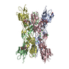
Mass: 122.143 Da / Num. of mol.: 1 / Source method: obtained synthetically / Formula: C4H12NO3 / Comment: pH buffer*YM
#4: Chemical
ChemComp-CA / CALCIUM ION


Mass: 40.078 Da / Num. of mol.: 14 / Source method: obtained synthetically / Formula: Ca
#5: Chemical
ChemComp-CL / CHLORIDE ION


Mass: 35.453 Da / Num. of mol.: 6 / Source method: obtained synthetically / Formula: Cl
#6: Chemical ChemComp-NA / SODIUM ION


Mass: 22.990 Da / Num. of mol.: 2 / Source method: obtained synthetically / Formula: Na
#7: Chemical ChemComp-IOD / IODIDE ION


Mass: 126.904 Da / Num. of mol.: 1 / Source method: obtained synthetically / Formula: I
#8: Water ChemComp-HOH / water


Mass: 18.015 Da / Num. of mol.: 33 / Source method: isolated from a natural source / Formula: H2O

-
Details

Has ligand of interestN

-
Experimental details

-
Experiment

ExperimentMethod: X-RAY DIFFRACTION / Number of used crystals: 1

-
Sample preparation

CrystalDensity Matthews: 2.71 Å3/Da / Density % sol: 54.66 %
Crystal growTemperature: 277 K / Method: vapor diffusion, sitting drop / pH: 8 / Details: 0.2 MM NaI 20% PEG3350 CDHR5 EC1-2

-
Data collection

DiffractionMean temperature: 100 K / Serial crystal experiment: N
Diffraction sourceSource: SYNCHROTRON / Site: APS / Beamline: 24-ID-E / Wavelength: 0.97918 Å
DetectorType: DECTRIS EIGER X 16M / Detector: PIXEL / Date: Dec 1, 2020
RadiationProtocol: SINGLE WAVELENGTH / Monochromatic (M) / Laue (L): M / Scattering type: x-ray
Radiation wavelengthWavelength: 0.97918 Å / Relative weight: 1
ReflectionResolution: 3.175→50 Å / Num. obs: 18809 / % possible obs: 96.3 % / Redundancy: 3.5 % / CC1/2: 0.987 / Net I/σ(I): 5.6
Reflection shellResolution: 3.175→3.23 Å / Redundancy: 2.5 % / Mean I/σ(I) obs: 2.63 / Num. unique obs: 810 / CC1/2: 0.277 / % possible all: 89.1

-
Processing

Software
NameVersionClassification
REFMAC5.8.0267refinement
HKL-2000data reduction
HKL-2000data scaling
PHASERphasing
RefinementMethod to determine structure: MOLECULAR REPLACEMENT
Starting model: 5CZR
Resolution: 3.175→49.147 Å / Cor.coef. Fo:Fc: 0.877 / Cor.coef. Fo:Fc free: 0.81 / SU B: 25.538 / SU ML: 0.438 / Cross valid method: FREE R-VALUE / ESU R Free: 0.593
Details: Hydrogens have been added in their riding positions
RfactorNum. reflection% reflection
Rfree0.2896 862 4.767 %
Rwork0.2135 17221 -
all0.217 --
obs-18083 95.219 %
Solvent computationIon probe radii: 0.8 Å / Shrinkage radii: 0.8 Å / VDW probe radii: 1.2 Å / Solvent model: MASK BULK SOLVENT
Displacement parametersBiso mean: 41.168 Å2
Baniso -1Baniso -2Baniso -3
1--0.375 Å2-0 Å2-1.706 Å2
2--0.913 Å20 Å2
3---0.25 Å2
Refinement stepCycle: LAST / Resolution: 3.175→49.147 Å
ProteinNucleic acidLigandSolventTotal
Num. atoms6500 0 187 33 6720
Refine LS restraints
Refine-IDTypeDev idealDev ideal targetNumber
X-RAY DIFFRACTIONr_bond_refined_d0.0170.0136817
X-RAY DIFFRACTIONr_bond_other_d0.0040.0176366
X-RAY DIFFRACTIONr_angle_refined_deg2.021.6659295
X-RAY DIFFRACTIONr_angle_other_deg1.4891.59814703
X-RAY DIFFRACTIONr_dihedral_angle_1_deg7.2315841
X-RAY DIFFRACTIONr_dihedral_angle_2_deg37.53125.069290
X-RAY DIFFRACTIONr_dihedral_angle_3_deg19.808151054
X-RAY DIFFRACTIONr_dihedral_angle_4_deg15.7861512
X-RAY DIFFRACTIONr_chiral_restr0.0730.2942
X-RAY DIFFRACTIONr_gen_planes_refined0.0090.027607
X-RAY DIFFRACTIONr_gen_planes_other0.0030.021441
X-RAY DIFFRACTIONr_nbd_refined0.2440.21069
X-RAY DIFFRACTIONr_symmetry_nbd_other0.2260.25797
X-RAY DIFFRACTIONr_nbtor_refined0.1850.23223
X-RAY DIFFRACTIONr_symmetry_nbtor_other0.1080.23763
X-RAY DIFFRACTIONr_xyhbond_nbd_refined0.1530.2116
X-RAY DIFFRACTIONr_symmetry_xyhbond_nbd_other0.1370.22
X-RAY DIFFRACTIONr_metal_ion_refined0.3130.253
X-RAY DIFFRACTIONr_symmetry_nbd_refined0.2970.224
X-RAY DIFFRACTIONr_nbd_other0.3230.256
X-RAY DIFFRACTIONr_symmetry_xyhbond_nbd_refined0.2660.21
X-RAY DIFFRACTIONr_mcbond_it5.4693.9923386
X-RAY DIFFRACTIONr_mcbond_other5.4673.9913385
X-RAY DIFFRACTIONr_mcangle_it8.4665.9794219
X-RAY DIFFRACTIONr_mcangle_other8.4655.984220
X-RAY DIFFRACTIONr_scbond_it7.7664.7463431
X-RAY DIFFRACTIONr_scbond_other7.7664.7463431
X-RAY DIFFRACTIONr_scangle_it11.3866.8495076
X-RAY DIFFRACTIONr_scangle_other11.3856.8515077
X-RAY DIFFRACTIONr_lrange_it15.89979.89326635
X-RAY DIFFRACTIONr_lrange_other15.89979.89526630
Refine LS restraints NCS

Refine-ID: X-RAY DIFFRACTION / Type: tight positional; tight thermal / Weight Biso : 0.86601 / Weight position: 0.0866

Ens-IDDom-IDAuth asym-IDRms dev Biso 2)Rms dev position (Å)
11A8.866220.50982
12B8.866220.50982
23A8.281940.63083
24C8.281940.63083
35A7.25430.4381
36D7.25430.4381
47B7.614330.45163
48C7.614330.45163
59B8.546550.56262
510D8.546550.56262
611C8.117020.57889
612D8.117020.57889
LS refinement shell
Resolution (Å)Rfactor RfreeNum. reflection RfreeRfactor RworkNum. reflection RworkRefine-ID% reflection obs (%)
3.175-3.2570.313570.2351008X-RAY DIFFRACTION77.4545
3.257-3.3460.328710.2331195X-RAY DIFFRACTION92.6794
3.346-3.4430.338660.2351156X-RAY DIFFRACTION93.2824
3.443-3.5490.341750.2321160X-RAY DIFFRACTION94.8541
3.549-3.6650.336540.2311131X-RAY DIFFRACTION96.1851
3.665-3.7930.312520.2141134X-RAY DIFFRACTION97.5329
3.793-3.9360.334460.1881099X-RAY DIFFRACTION98.0308
3.936-4.0970.244320.1851061X-RAY DIFFRACTION98.2914
4.097-4.2790.235460.1771008X-RAY DIFFRACTION97.0534
4.279-4.4870.244420.179947X-RAY DIFFRACTION97.1513
4.487-4.7290.199410.161881X-RAY DIFFRACTION94.4672
4.729-5.0160.208420.152887X-RAY DIFFRACTION98.62
5.016-5.3610.243470.166827X-RAY DIFFRACTION99.3182
5.361-5.7890.298370.186779X-RAY DIFFRACTION99.5122
5.789-6.340.283390.209697X-RAY DIFFRACTION99.0579
6.34-7.0850.302290.226653X-RAY DIFFRACTION98.8406
7.085-8.1740.281290.225555X-RAY DIFFRACTION98.1513
8.174-9.9950.293250.299464X-RAY DIFFRACTION94.9515
9.995-14.0680.363240.334372X-RAY DIFFRACTION97.0588
14.068-49.1470.39480.415207X-RAY DIFFRACTION90.7173

+
About Yorodumi

-
News

-
Feb 9, 2022. New format data for meta-information of EMDB entries

New format data for meta-information of EMDB entries

  • Version 3 of the EMDB header file is now the official format.
  • The previous official version 1.9 will be removed from the archive.

Related info.:EMDB header

External links:wwPDB to switch to version 3 of the EMDB data model

-
Aug 12, 2020. Covid-19 info

Covid-19 info

URL: https://pdbjlvh1.pdbj.org/emnavi/covid19.php

New page: Covid-19 featured information page in EM Navigator.

Related info.:Covid-19 info / Mar 5, 2020. Novel coronavirus structure data

+
Mar 5, 2020. Novel coronavirus structure data

Novel coronavirus structure data

Related info.:Yorodumi Speices / Aug 12, 2020. Covid-19 info

External links:COVID-19 featured content - PDBj / Molecule of the Month (242):Coronavirus Proteases

+
Jan 31, 2019. EMDB accession codes are about to change! (news from PDBe EMDB page)

EMDB accession codes are about to change! (news from PDBe EMDB page)

  • The allocation of 4 digits for EMDB accession codes will soon come to an end. Whilst these codes will remain in use, new EMDB accession codes will include an additional digit and will expand incrementally as the available range of codes is exhausted. The current 4-digit format prefixed with “EMD-” (i.e. EMD-XXXX) will advance to a 5-digit format (i.e. EMD-XXXXX), and so on. It is currently estimated that the 4-digit codes will be depleted around Spring 2019, at which point the 5-digit format will come into force.
  • The EM Navigator/Yorodumi systems omit the EMD- prefix.

Related info.:Q: What is EMD? / ID/Accession-code notation in Yorodumi/EM Navigator

External links:EMDB Accession Codes are Changing Soon! / Contact to PDBj

+
Jul 12, 2017. Major update of PDB

Major update of PDB

  • wwPDB released updated PDB data conforming to the new PDBx/mmCIF dictionary.
  • This is a major update changing the version number from 4 to 5, and with Remediation, in which all the entries are updated.
  • In this update, many items about electron microscopy experimental information are reorganized (e.g. em_software).
  • Now, EM Navigator and Yorodumi are based on the updated data.

External links:wwPDB Remediation / Enriched Model Files Conforming to OneDep Data Standards Now Available in the PDB FTP Archive

-
Yorodumi

Thousand views of thousand structures

  • Yorodumi is a browser for structure data from EMDB, PDB, SASBDB, etc.
  • This page is also the successor to EM Navigator detail page, and also detail information page/front-end page for Omokage search.
  • The word "yorodu" (or yorozu) is an old Japanese word meaning "ten thousand". "mi" (miru) is to see.

Related info.:EMDB / PDB / SASBDB / Comparison of 3 databanks / Yorodumi Search / Aug 31, 2016. New EM Navigator & Yorodumi / Yorodumi Papers / Jmol/JSmol / Function and homology information / Changes in new EM Navigator and Yorodumi

Read more