| Entry | Database: PDB / ID: 3zdo
|
|---|
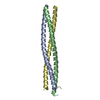
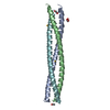
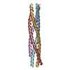
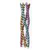
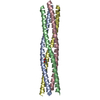
| Title | Tetramerization domain of Measles virus phosphoprotein |
|---|
 Components Components | PHOSPHOPROTEIN |
|---|
 Keywords Keywords | VIRAL PROTEIN / COILED-COIL |
|---|
| Function / homology |  Function and homology information Function and homology information
viral genome replication / RNA-directed RNA polymerase activity / DNA-templated transcription / RNA binding / metal ion bindingSimilarity search - Function RNA polymerase, phosphoprotein P, C-terminal XD, paramyxovirinae / Single alpha-helices involved in coiled-coils or other helix-helix interfaces - #110 / Paramyxovirus structural protein P/V, N-terminal domain / Paramyxovirus structural protein V/P N-terminus / P/V phosphoprotein, paramyxoviral / Paramyxovirus P/V phosphoprotein C-terminal / Single alpha-helices involved in coiled-coils or other helix-helix interfaces / Up-down Bundle / Mainly AlphaSimilarity search - Domain/homology |
|---|
| Biological species |   MEASLES VIRUS MEASLES VIRUS |
|---|
| Method |  X-RAY DIFFRACTION / SYNCHROTRON / MOLECULAR REPLACEMENT / Resolution: 2.07 Å X-RAY DIFFRACTION / SYNCHROTRON / MOLECULAR REPLACEMENT / Resolution: 2.07 Å |
|---|
 Authors Authors | Communie, G. / Crepin, T. / Jensen, M.R. / Blackledge, M. / Ruigrok, R.W.H. |
|---|
 Citation Citation | Journal: J.Virol. / Year: 2013
Title: Structure of the Tetramerization Domain of Measles Virus Phosphoprotein.
Authors: Communie, G. / Crepin, T. / Maurin, D. / Ringkjobing Jensen, M. / Blackledge, M. / Ruigrok, R.W. |
|---|
| History | | Deposition | Nov 29, 2012 | Deposition site: PDBE / Processing site: PDBE |
|---|
| Revision 1.0 | Apr 24, 2013 | Provider: repository / Type: Initial release |
|---|
| Revision 1.1 | Jun 12, 2013 | Group: Database references |
|---|
| Revision 1.2 | May 1, 2024 | Group: Data collection / Database references ...Data collection / Database references / Derived calculations / Other / Refinement description
Category: chem_comp_atom / chem_comp_bond ...chem_comp_atom / chem_comp_bond / database_2 / pdbx_database_status / pdbx_initial_refinement_model / struct_site
Item: _database_2.pdbx_DOI / _database_2.pdbx_database_accession ..._database_2.pdbx_DOI / _database_2.pdbx_database_accession / _pdbx_database_status.status_code_sf / _struct_site.pdbx_auth_asym_id / _struct_site.pdbx_auth_comp_id / _struct_site.pdbx_auth_seq_id |
|---|
|
|---|
 Movie
Movie Controller


 Open data
 Basic information
 Structure visualization
 Downloads & links
 Other downloads

 PDBj
PDBj
 Assembly
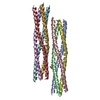


 Mass: 118.174 Da / Num. of mol.: 1 / Source method: obtained synthetically / Formula: C6H14O2 / Comment: precipitant*YM
 Mass: 18.015 Da / Num. of mol.: 92 / Source method: isolated from a natural source / Formula: H2O
 Sample preparation
 / Beamline: ID23-2 / Wavelength: 0.8726
  Processing