muscle cell apoptotic process / regulation of skeletal muscle contraction by calcium ion signaling / regulation of myotube differentiation / myosin phosphatase regulator activity / regulation of synapse structural plasticity / nuclear envelope organization / regulation of heart contraction / nuclear outer membrane / Ion homeostasis / sarcoplasmic reticulum membrane ...muscle cell apoptotic process / regulation of skeletal muscle contraction by calcium ion signaling / regulation of myotube differentiation / myosin phosphatase regulator activity / regulation of synapse structural plasticity / nuclear envelope organization / regulation of heart contraction / nuclear outer membrane / Ion homeostasis / sarcoplasmic reticulum membrane / intracellular calcium ion homeostasis / nuclear membrane / protein phosphorylation / mitochondrial outer membrane / non-specific serine/threonine protein kinase / protein serine kinase activity / protein serine/threonine kinase activity / endoplasmic reticulum membrane / ATP binding / metal ion binding / plasma membrane / cytosol Similarity search - Function
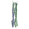
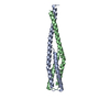
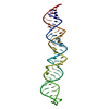
Myotonic dystrophy protein kinase, coiled coil / DMPK coiled coil domain like / Single alpha-helices involved in coiled-coils or other helix-helix interfaces - #340 / : / Extension to Ser/Thr-type protein kinases / AGC-kinase, C-terminal / AGC-kinase C-terminal domain profile. / Single alpha-helices involved in coiled-coils or other helix-helix interfaces / Serine/threonine-protein kinase, active site / Serine/Threonine protein kinases active-site signature. ...Myotonic dystrophy protein kinase, coiled coil / DMPK coiled coil domain like / Single alpha-helices involved in coiled-coils or other helix-helix interfaces - #340 / : / Extension to Ser/Thr-type protein kinases / AGC-kinase, C-terminal / AGC-kinase C-terminal domain profile. / Single alpha-helices involved in coiled-coils or other helix-helix interfaces / Serine/threonine-protein kinase, active site / Serine/Threonine protein kinases active-site signature. / Protein kinase domain / Serine/Threonine protein kinases, catalytic domain / Protein kinase, ATP binding site / Protein kinases ATP-binding region signature. / Protein kinase domain profile. / Protein kinase domain / Protein kinase-like domain superfamily / Up-down Bundle / Mainly Alpha Similarity search - Domain/homology
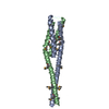
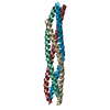
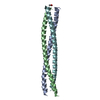
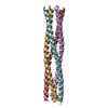
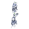
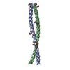
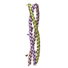
#1: Journal: ACTA CRYSTALLOGR.,SECT.D / Year: 2004 Title: Crystallization and preliminary X-ray analysis of the coiled-coil domain of dystrophia myotonica kinase Authors: Garcia, P. / Marino, M. / Mayans, O.
In the structure databanks used in Yorodumi, some data are registered as the other names, "COVID-19 virus" and "2019-nCoV". Here are the details of the virus and the list of structure data.
Jan 31, 2019. EMDB accession codes are about to change! (news from PDBe EMDB page)
EMDB accession codes are about to change! (news from PDBe EMDB page)
The allocation of 4 digits for EMDB accession codes will soon come to an end. Whilst these codes will remain in use, new EMDB accession codes will include an additional digit and will expand incrementally as the available range of codes is exhausted. The current 4-digit format prefixed with “EMD-” (i.e. EMD-XXXX) will advance to a 5-digit format (i.e. EMD-XXXXX), and so on. It is currently estimated that the 4-digit codes will be depleted around Spring 2019, at which point the 5-digit format will come into force.
The EM Navigator/Yorodumi systems omit the EMD- prefix.
Related info.:Q: What is EMD? / ID/Accession-code notation in Yorodumi/EM Navigator
Yorodumi is a browser for structure data from EMDB, PDB, SASBDB, etc.
This page is also the successor to EM Navigator detail page, and also detail information page/front-end page for Omokage search.
The word "yorodu" (or yorozu) is an old Japanese word meaning "ten thousand". "mi" (miru) is to see.
Related info.:EMDB / PDB / SASBDB / Comparison of 3 databanks / Yorodumi Search / Aug 31, 2016. New EM Navigator & Yorodumi / Yorodumi Papers / Jmol/JSmol / Function and homology information / Changes in new EM Navigator and Yorodumi
 Movie
Movie Controller


 Open data
 Basic information
 Components
 Keywords
 Function and homology information
 Homo sapiens (human)
 X-RAY DIFFRACTION /
  Authors
 Citation
 Structure visualization
 Downloads & links
 Other downloads
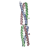

 PDBj
PDBj


 Assembly
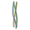

 Mass: 18.015 Da / Num. of mol.: 295 / Source method: isolated from a natural source / Formula: H2O
 Sample preparation
 / Beamline: ID14-2 / Wavelength: 0.98, 0.9793, 0.9795, 0.9392
 Processing