 Movie
Movie Controller

[English] 日本語
 Yorodumi
- PDB-1gu4: Crystal structure of C/EBPBETA BZIP homodimer bound to a high aff... -
+ Open data

- Basic information
| Entry | Database: PDB / ID: 1gu4 | ||||||
|---|---|---|---|---|---|---|---|
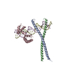
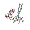
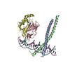
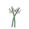
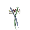
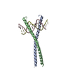
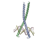
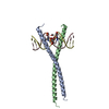
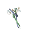
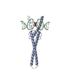
| Title | Crystal structure of C/EBPBETA BZIP homodimer bound to a high affinity DNA fragment | ||||||
 Components Components |
| ||||||
 Keywords Keywords | TRANSCRIPTION/DNA / PROTEIN-DNA COMPLEX / TRANSCRIPTION FACTOR / BZIP / C/EBP / HYPOTHETICAL PROTEIN / TRANSCRIPTION-DNA complex | ||||||
| Function / homology |  Function and homology information Function and homology informationregulation of odontoblast differentiation / C/EBP complex / positive regulation of sodium-dependent phosphate transport / CHOP-C/EBP complex / positive regulation of biomineral tissue development / granuloma formation / myeloid cell development / integrated stress response signaling / T-helper 1 cell activation / hepatocyte proliferation ...regulation of odontoblast differentiation / C/EBP complex / positive regulation of sodium-dependent phosphate transport / CHOP-C/EBP complex / positive regulation of biomineral tissue development / granuloma formation / myeloid cell development / integrated stress response signaling / T-helper 1 cell activation / hepatocyte proliferation / Response of EIF2AK1 (HRI) to heme deficiency / ATF4 activates genes in response to endoplasmic reticulum stress / nuclear glucocorticoid receptor binding / mammary gland epithelial cell differentiation / regulation of osteoclast differentiation / condensed chromosome, centromeric region / regulation of dendritic cell differentiation / regulation of interleukin-6 production / mammary gland epithelial cell proliferation / histone acetyltransferase binding / positive regulation of interleukin-4 production / regulation of cell differentiation / Transcriptional Regulation by VENTX / ubiquitin-like protein ligase binding / intrinsic apoptotic signaling pathway in response to endoplasmic reticulum stress / cellular response to interleukin-1 / positive regulation of osteoblast differentiation / embryonic placenta development / Response of EIF2AK4 (GCN2) to amino acid deficiency / positive regulation of fat cell differentiation / RNA polymerase II core promoter sequence-specific DNA binding / ovarian follicle development / negative regulation of T cell proliferation / Nuclear events stimulated by ALK signaling in cancer / liver regeneration / brown fat cell differentiation / response to endoplasmic reticulum stress / acute-phase response / RNA polymerase II transcription regulatory region sequence-specific DNA binding / cellular response to amino acid stimulus / Transcriptional regulation of white adipocyte differentiation / chromatin DNA binding / DNA-binding transcription repressor activity, RNA polymerase II-specific / RNA polymerase II transcription regulator complex / histone deacetylase binding / kinase binding / Transcriptional regulation of granulopoiesis / neuron differentiation / nuclear matrix / memory / sequence-specific double-stranded DNA binding / positive regulation of inflammatory response / positive regulation of cold-induced thermogenesis / cellular response to lipopolysaccharide / Senescence-Associated Secretory Phenotype (SASP) / DNA-binding transcription activator activity, RNA polymerase II-specific / transcription by RNA polymerase II / negative regulation of neuron apoptotic process / DNA-binding transcription factor activity, RNA polymerase II-specific / defense response to bacterium / transcription cis-regulatory region binding / immune response / RNA polymerase II cis-regulatory region sequence-specific DNA binding / DNA-binding transcription factor activity / inflammatory response / protein heterodimerization activity / regulation of transcription by RNA polymerase II / regulation of DNA-templated transcription / positive regulation of DNA-templated transcription / chromatin / negative regulation of transcription by RNA polymerase II / protein homodimerization activity / positive regulation of transcription by RNA polymerase II / DNA binding / nucleoplasm / nucleus / cytoplasm Similarity search - Function | ||||||
| Biological species |  HOMO SAPIENS (human) HOMO SAPIENS (human)SYNTHETIC CONSTRUCT (others) | ||||||
| Method |  X-RAY DIFFRACTION / SYNCHROTRON / MOLECULAR REPLACEMENT / Resolution: 1.8 Å X-RAY DIFFRACTION / SYNCHROTRON / MOLECULAR REPLACEMENT / Resolution: 1.8 Å | ||||||
 Authors Authors | Tahirov, T.H. / Ogata, K. | ||||||
 Citation Citation | Journal: To be Published Title: Structural Basis for Flexible Base Recognition by C/Ebpbeta Authors: Tahirov, T.H. / Inoue-Bungo, T. / Sato, K. / Sasaki, M. / Ogata, K. #1: Journal: Cell (Cambridge,Mass.) / Year: 2002Title: Mechanism of C-Myb-C/Ebpbeta Cooperation from Separated Sites on a Promoter Authors: Tahirov, T.H. / Sato, K. / Ichikawa-Iwata, E. / Sasaki, M. / Inoue-Bungo, T. / Shiina, M. / Kimura, K. / Takata, S. / Fujikawa, A. / Morii, H. / Kumasaka, T. / Yamamoto, M. / Ishii, S. / Ogata, K. #2: Journal: Acta Crystallogr.,Sect.D / Year: 2001 Title: Crystallization and Preliminary X-Ray Analysis of the C/Ebpbeta C-Terminal Region in Complex with DNA Authors: Tahirov, T.H. / Inoue-Bungo, T. / Sasaki, M. / Fujikawa, A. / Kimura, K. / Sato, K. / Adachi, S. / Kamiyaogata, K. #3: Journal: Cell (Cambridge,Mass.) / Year: 2001Title: Structural Analyses of DNA Recognition by the Aml1/ Runx-1 Runt Domain and its Allosteric Control by Cbfbeta Authors: Tahirov, T.H. / Inoue-Bungo, T. / Morii, H. / Fujikawa, A. / Sasaki, M. / Kimura, K. / Shiina, M. / Sato, K. / Kumasaka, T. / Yamamoto, M. / Ishii, S. / Ogata, K. #4: Journal: Acta Crystallogr.,Sect.D / Year: 2001 Title: Crystallization and Preliminary X-Ray Analyses of Quaternary, Ternary and Binary Protein-DNA Complexes with Involvement of Aml1/Runx-1/Cbfalpha Runt Domain, Cbfbeta and the C/Ebpbeta bZIP Region Authors: Tahirov, T.H. / Inoue, T. / Sasaki, M. / Shiina, M. / Kimura, K. / Sato, K. / Kumasaka, T. / Yamamoto, M. / Kamiya, N. / Ogata, K. #5: Journal: Acta Crystallogr.,Sect.D / Year: 2001 Title: Crystals of Ternary Protein-DNA Complexes Composed of DNA-Binding Domains C-Myb or V-Myb, C/Ebpalpha or C/Ebpbeta and Tom-1A Promoter Fragment Authors: Tahirov, T.H. / Inoue, T. / Sasaki, M. / Shiina, M. / Kimura, K. / Sato, K. / Kumasaka, T. / Yamamoto, M. / Kamiya, N. / Ogata, K. | ||||||
| History |
|
- Structure visualization
| Structure viewer | Molecule: MolmilJmol/JSmol |
|---|
- Downloads & links
-Download
| PDBx/mmCIF format | 1gu4.cif.gz | 67.2 KB | Display | PDBx/mmCIF format |
|---|---|---|---|---|
| PDB format | pdb1gu4.ent.gz | 47.6 KB | Display | PDB format |
| PDBx/mmJSON format | 1gu4.json.gz | Tree view | PDBx/mmJSON format | |
| Others |  Other downloads Other downloads |
-Validation report
| Arichive directory | https://data.pdbj.org/pub/pdb/validation_reports/gu/1gu4ftp://data.pdbj.org/pub/pdb/validation_reports/gu/1gu4 | HTTPS FTP |
|---|
-Related structure data
| Related structure data | 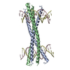 1gtwSC 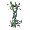 1gu5C  2e42C 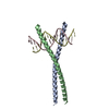 2e43C S: Starting model for refinement C: citing same article ( |
|---|---|
| Similar structure data |


-Links
 PDBj
PDBj


- Assembly
| Deposited unit | 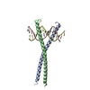
| ||||||||
|---|---|---|---|---|---|---|---|---|---|
| 1 | 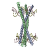
| ||||||||
| Unit cell |
| ||||||||
| Components on special symmetry positions |
|
-Components
| #1: Protein | Mass: 9381.868 Da / Num. of mol.: 2 / Fragment: BZIP DOMAIN, RESIDUES 259-336 Source method: isolated from a genetically manipulated source Source: (gene. exp.) HOMO SAPIENS (human) / Plasmid: PAR2156 / Production host:  #2: DNA chain | | Mass: 4937.228 Da / Num. of mol.: 1 / Source method: obtained synthetically / Source: (synth.) SYNTHETIC CONSTRUCT (others) #3: DNA chain | | Mass: 4857.179 Da / Num. of mol.: 1 / Source method: obtained synthetically / Source: (synth.) SYNTHETIC CONSTRUCT (others) #4: Water | ChemComp-HOH / |  Mass: 18.015 Da / Num. of mol.: 311 / Source method: isolated from a natural source / Formula: H2O Mass: 18.015 Da / Num. of mol.: 311 / Source method: isolated from a natural source / Formula: H2O |
|---|
-Experimental details
-Experiment
| Experiment | Method: X-RAY DIFFRACTION / Number of used crystals: 1 |
|---|
- Sample preparation
| Crystal | Density Matthews: 3.69 Å3/Da / Density % sol: 66 % |
|---|---|
| Crystal grow | pH: 6 Details: 0.2 M POTASSIUM CHLORIDE, 0.01 M MAGNESIUM SULFATE, 10.0% V/V PEG 400, 0.05 M MES BUFFER PH 6.0, PROTEIN-DNA COMPLEX CONCENTRATION WAS 12 MG/ML AND CONTAINS 0.01 M DTT, PROTEIN:DNA RATIO WAS ...Details: 0.2 M POTASSIUM CHLORIDE, 0.01 M MAGNESIUM SULFATE, 10.0% V/V PEG 400, 0.05 M MES BUFFER PH 6.0, PROTEIN-DNA COMPLEX CONCENTRATION WAS 12 MG/ML AND CONTAINS 0.01 M DTT, PROTEIN:DNA RATIO WAS 1:1.2. FOR CRYOPROTECTION THE CONCENTRATION OF PEG 400 WAS ADJUSTED TO 36% V/V |
-Data collection
| Diffraction | Mean temperature: 90 K |
|---|---|
| Diffraction source | Source: SYNCHROTRON / Site: SPring-8  / Beamline: BL45XU / Wavelength: 1.02 / Beamline: BL45XU / Wavelength: 1.02 |
| Detector | Type: RIGAKU RAXIS V / Detector: IMAGE PLATE / Date: Oct 12, 2001 / Details: MIRRORS |
| Radiation | Monochromator: DIAMOND / Protocol: SINGLE WAVELENGTH / Monochromatic (M) / Laue (L): M / Scattering type: x-ray |
| Radiation wavelength | Wavelength: 1.02 Å / Relative weight: 1 |
| Reflection | Resolution: 1.75→30 Å / Num. obs: 42716 / % possible obs: 99.4 % / Observed criterion σ(I): 0 / Redundancy: 5.452 % / Biso Wilson estimate: 31.9 Å2 / Rmerge(I) obs: 0.094 / Net I/σ(I): 21.7331 |
| Reflection shell | Resolution: 1.75→1.78 Å / Redundancy: 3.96 % / Rmerge(I) obs: 0.455 / Mean I/σ(I) obs: 2.079 / % possible all: 98.3 |
- Processing
| Software |
| ||||||||||||||||||||||||||||||||||||||||||||||||||||||||||||||||||||||||||||||||
|---|---|---|---|---|---|---|---|---|---|---|---|---|---|---|---|---|---|---|---|---|---|---|---|---|---|---|---|---|---|---|---|---|---|---|---|---|---|---|---|---|---|---|---|---|---|---|---|---|---|---|---|---|---|---|---|---|---|---|---|---|---|---|---|---|---|---|---|---|---|---|---|---|---|---|---|---|---|---|---|---|---|
| Refinement | Method to determine structure: MOLECULAR REPLACEMENT Starting model: PDB ENTRY 1GTW Resolution: 1.8→29.95 Å / Rfactor Rfree error: 0.006 / Data cutoff high absF: 1627019.3 / Isotropic thermal model: RESTRAINED / Cross valid method: THROUGHOUT / σ(F): 0
| ||||||||||||||||||||||||||||||||||||||||||||||||||||||||||||||||||||||||||||||||
| Solvent computation | Solvent model: FLAT MODEL / Bsol: 54.8637 Å2 / ksol: 0.348039 e/Å3 | ||||||||||||||||||||||||||||||||||||||||||||||||||||||||||||||||||||||||||||||||
| Displacement parameters | Biso mean: 39.9 Å2
| ||||||||||||||||||||||||||||||||||||||||||||||||||||||||||||||||||||||||||||||||
| Refine analyze |
| ||||||||||||||||||||||||||||||||||||||||||||||||||||||||||||||||||||||||||||||||
| Refinement step | Cycle: LAST / Resolution: 1.8→29.95 Å
| ||||||||||||||||||||||||||||||||||||||||||||||||||||||||||||||||||||||||||||||||
| Refine LS restraints |
| ||||||||||||||||||||||||||||||||||||||||||||||||||||||||||||||||||||||||||||||||
| LS refinement shell | Resolution: 1.8→1.91 Å / Rfactor Rfree error: 0.018 / Total num. of bins used: 6
| ||||||||||||||||||||||||||||||||||||||||||||||||||||||||||||||||||||||||||||||||
| Xplor file |
|
