| Entry | Database: PDB / ID: 7m6k
|
|---|
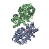
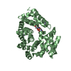
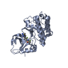
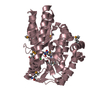
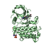
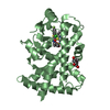
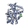
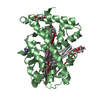
| Title | Crystal structure of the ARM domain from Drosophila SARM1 in complex with VMN |
|---|
 Components Components | Isoform B of NAD(+) hydrolase sarm1 |
|---|
 Keywords Keywords | IMMUNE SYSTEM / neurotoxicity / axon degeneration / NADase / ARM domain |
|---|
| Function / homology |  Function and homology information Function and homology information
positive regulation of receptor signaling pathway via STAT / NAD+ nucleosidase activity / NAD+ catabolic process / ADP-ribosyl cyclase/cyclic ADP-ribose hydrolase / NAD+ nucleosidase activity, cyclic ADP-ribose generating / STAT family protein binding / response to axon injury / signaling adaptor activity / antiviral innate immune response / defense response to virus ...positive regulation of receptor signaling pathway via STAT / NAD+ nucleosidase activity / NAD+ catabolic process / ADP-ribosyl cyclase/cyclic ADP-ribose hydrolase / NAD+ nucleosidase activity, cyclic ADP-ribose generating / STAT family protein binding / response to axon injury / signaling adaptor activity / antiviral innate immune response / defense response to virus / neuron projection / axon / neuronal cell body / dendrite / signal transduction / mitochondrion / cytosolSimilarity search - Function Sterile alpha and TIR motif-containing protein 1 / TIR domain / Toll - interleukin 1 - resistance / TIR domain profile. / Toll/interleukin-1 receptor homology (TIR) domain / Toll/interleukin-1 receptor homology (TIR) domain superfamily / SAM domain (Sterile alpha motif) / SAM domain profile. / Sterile alpha motif. / Sterile alpha motif domain ...Sterile alpha and TIR motif-containing protein 1 / TIR domain / Toll - interleukin 1 - resistance / TIR domain profile. / Toll/interleukin-1 receptor homology (TIR) domain / Toll/interleukin-1 receptor homology (TIR) domain superfamily / SAM domain (Sterile alpha motif) / SAM domain profile. / Sterile alpha motif. / Sterile alpha motif domain / Sterile alpha motif/pointed domain superfamily / Armadillo-like helical / Armadillo-type foldSimilarity search - Domain/homology |
|---|
| Biological species |   Drosophila melanogaster (fruit fly) Drosophila melanogaster (fruit fly) |
|---|
| Method |  X-RAY DIFFRACTION / SYNCHROTRON / MOLECULAR REPLACEMENT / Resolution: 1.69 Å X-RAY DIFFRACTION / SYNCHROTRON / MOLECULAR REPLACEMENT / Resolution: 1.69 Å |
|---|
 Authors Authors | Gu, W. / Luo, Z. / Kobe, B. |
|---|
| Funding support |  Australia, 2items Australia, 2items | Organization | Grant number | Country |
|---|
| National Health and Medical Research Council (NHMRC, Australia) | 1160570 | Australia | | Australian Research Council (ARC) | FL180100109 | Australia |
|
|---|
 Citation Citation | Journal: Elife / Year: 2021
Title: Neurotoxin-mediated potent activation of the axon degeneration regulator SARM1.
Authors: Loreto, A. / Angeletti, C. / Gu, W. / Osborne, A. / Nieuwenhuis, B. / Gilley, J. / Merlini, E. / Arthur-Farraj, P. / Amici, A. / Luo, Z. / Hartley-Tassell, L. / Ve, T. / Desrochers, L.M. / ...Authors: Loreto, A. / Angeletti, C. / Gu, W. / Osborne, A. / Nieuwenhuis, B. / Gilley, J. / Merlini, E. / Arthur-Farraj, P. / Amici, A. / Luo, Z. / Hartley-Tassell, L. / Ve, T. / Desrochers, L.M. / Wang, Q. / Kobe, B. / Orsomando, G. / Coleman, M.P. |
|---|
| History | | Deposition | Mar 25, 2021 | Deposition site: RCSB / Processing site: RCSB |
|---|
| Revision 1.0 | Feb 2, 2022 | Provider: repository / Type: Initial release |
|---|
| Revision 1.1 | Oct 18, 2023 | Group: Data collection / Refinement description
Category: chem_comp_atom / chem_comp_bond / pdbx_initial_refinement_model |
|---|
| Revision 1.2 | Oct 16, 2024 | Group: Structure summary / Category: pdbx_entry_details / pdbx_modification_feature / Item: _pdbx_entry_details.has_protein_modification |
|---|
|
|---|
 Movie
Movie Controller

 Yorodumi
 Open data
 Basic information
 Structure visualization
 Downloads & links
 Other downloads


 PDBj
PDBj
 Assembly