 Movie
Movie Controller

[English] 日本語
 Yorodumi
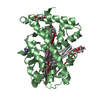
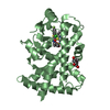
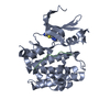
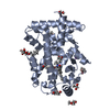
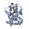
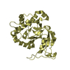
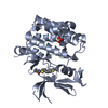
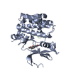
- PDB-7bq2: X-ray structure of human PPARalpha ligand binding domain-pemafibr... -
+ Open data

- Basic information
| Entry | Database: PDB / ID: 7bq2 | ||||||
|---|---|---|---|---|---|---|---|
| Title | X-ray structure of human PPARalpha ligand binding domain-pemafibrate-SRC1 coactivator peptide co-crystals obtained by soaking | ||||||
 Components Components |
| ||||||
 Keywords Keywords | TRANSCRIPTION / Nuclear receptor / Protein-ligand complex / PPAR | ||||||
| Function / homology |  Function and homology information Function and homology informationregulation of fatty acid transport / enamel mineralization / positive regulation of fatty acid beta-oxidation / regulation of ketone metabolic process / cellular response to fructose stimulus / negative regulation of hepatocyte apoptotic process / negative regulation of cell growth involved in cardiac muscle cell development / regulation of fatty acid metabolic process / positive regulation of fatty acid oxidation / behavioral response to nicotine ...regulation of fatty acid transport / enamel mineralization / positive regulation of fatty acid beta-oxidation / regulation of ketone metabolic process / cellular response to fructose stimulus / negative regulation of hepatocyte apoptotic process / negative regulation of cell growth involved in cardiac muscle cell development / regulation of fatty acid metabolic process / positive regulation of fatty acid oxidation / behavioral response to nicotine / negative regulation of appetite / lipoprotein metabolic process / negative regulation of leukocyte cell-cell adhesion / labyrinthine layer morphogenesis / regulation of thyroid hormone receptor signaling pathway / positive regulation of transcription from RNA polymerase II promoter by galactose / positive regulation of female receptivity / mitogen-activated protein kinase kinase kinase binding / ubiquitin conjugating enzyme binding / negative regulation of glycolytic process / NR1H2 & NR1H3 regulate gene expression to control bile acid homeostasis / positive regulation of fatty acid metabolic process / NFAT protein binding / DNA-binding transcription activator activity / nuclear steroid receptor activity / male mating behavior / hypothalamus development / negative regulation of cholesterol storage / epidermis development / progesterone receptor signaling pathway / cellular response to Thyroglobulin triiodothyronine / Synthesis of bile acids and bile salts / positive regulation of ATP biosynthetic process / negative regulation of macrophage derived foam cell differentiation / Synthesis of bile acids and bile salts via 27-hydroxycholesterol / Endogenous sterols / Synthesis of bile acids and bile salts via 7alpha-hydroxycholesterol / response to retinoic acid / protein-lysine-acetyltransferase activity / positive regulation of lipid biosynthetic process / nuclear retinoid X receptor binding / estrous cycle / phosphatase binding / Transcriptional regulation of brown and beige adipocyte differentiation by EBF2 / Recycling of bile acids and salts / histone acetyltransferase / NR1H3 & NR1H2 regulate gene expression linked to cholesterol transport and efflux / cellular response to hormone stimulus / estrogen receptor signaling pathway / intracellular receptor signaling pathway / nitric oxide metabolic process / negative regulation of reactive oxygen species biosynthetic process / peroxisome proliferator activated receptor signaling pathway / hormone-mediated signaling pathway / Regulation of lipid metabolism by PPARalpha / negative regulation of blood pressure / positive regulation of adipose tissue development / response to progesterone / lactation / BMAL1:CLOCK,NPAS2 activates circadian expression / regulation of cellular response to insulin stimulus / positive regulation of neuron differentiation / negative regulation of cytokine production involved in inflammatory response / response to nutrient / RORA,B,C and NR1D1 (REV-ERBA) regulate gene expression / Activation of gene expression by SREBF (SREBP) / SUMOylation of transcription cofactors / Expression of BMAL (ARNTL), CLOCK, and NPAS2 / MDM2/MDM4 family protein binding / negative regulation of miRNA transcription / positive regulation of gluconeogenesis / cerebellum development / fatty acid metabolic process / nuclear estrogen receptor binding / nuclear receptor binding / negative regulation of phosphatidylinositol 3-kinase/protein kinase B signal transduction / cellular response to starvation / gluconeogenesis / hippocampus development / SUMOylation of intracellular receptors / RNA polymerase II transcription regulatory region sequence-specific DNA binding / circadian regulation of gene expression / wound healing / Heme signaling / negative regulation of transforming growth factor beta receptor signaling pathway / PPARA activates gene expression / Transcriptional activation of mitochondrial biogenesis / Cytoprotection by HMOX1 / cerebral cortex development / Nuclear Receptor transcription pathway / male gonad development / Transcriptional regulation of white adipocyte differentiation / response to insulin / regulation of circadian rhythm / negative regulation of inflammatory response / DNA-binding transcription repressor activity, RNA polymerase II-specific / mRNA transcription by RNA polymerase II / nuclear receptor activity / RNA polymerase II transcription regulator complex / transcription coactivator binding Similarity search - Function | ||||||
| Biological species |  Homo sapiens (human) Homo sapiens (human) | ||||||
| Method |  X-RAY DIFFRACTION / SYNCHROTRON / MOLECULAR REPLACEMENT / molecular replacement / Resolution: 1.52 Å X-RAY DIFFRACTION / SYNCHROTRON / MOLECULAR REPLACEMENT / molecular replacement / Resolution: 1.52 Å | ||||||
 Authors Authors | Kamata, S. / Ishikawa, R. / Akahane, M. / Oyama, T. / Ishii, I. | ||||||
| Funding support |  Japan, 1items Japan, 1items
| ||||||
 Citation Citation | Journal: Iscience / Year: 2020 Title: PPAR alpha Ligand-Binding Domain Structures with Endogenous Fatty Acids and Fibrates. Authors: Kamata, S. / Oyama, T. / Saito, K. / Honda, A. / Yamamoto, Y. / Suda, K. / Ishikawa, R. / Itoh, T. / Watanabe, Y. / Shibata, T. / Uchida, K. / Suematsu, M. / Ishii, I. | ||||||
| History |
|
- Structure visualization
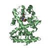
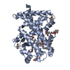
| Structure viewer | Molecule: MolmilJmol/JSmol |
|---|
- Downloads & links
-Download
| PDBx/mmCIF format | 7bq2.cif.gz | 76.3 KB | Display | PDBx/mmCIF format |
|---|---|---|---|---|
| PDB format | pdb7bq2.ent.gz | 53.7 KB | Display | PDB format |
| PDBx/mmJSON format | 7bq2.json.gz | Tree view | PDBx/mmJSON format | |
| Others |  Other downloads Other downloads |
-Validation report
| Arichive directory | https://data.pdbj.org/pub/pdb/validation_reports/bq/7bq2ftp://data.pdbj.org/pub/pdb/validation_reports/bq/7bq2 | HTTPS FTP |
|---|
-Related structure data
| Related structure data |  6kaxC 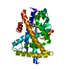 6kayC 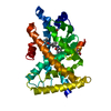 6kazC 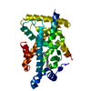 6kb0C 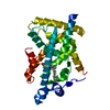 6kb1C 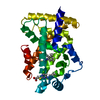 6kb2C 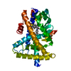 6kb3C 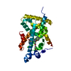 6kb4C 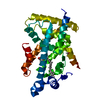 6kb5C 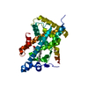 6kb6C 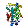 6kb7C 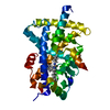 6kb8C 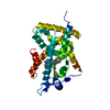 6kb9C  6kbaC 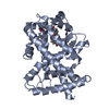 6kypC 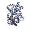 6l36C 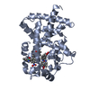 6l37C 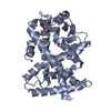 6l38C  6lx4C  6lx5C  6lx6C  6lx7C 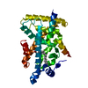 6lx8C  6lx9C 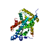 6lxaC  6lxbC  6lxcC  7bpyC  7bpzC  7bq0C  7bq1C  7bq3C  7bq4C 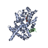 3sp6S S: Starting model for refinement C: citing same article ( |
|---|---|
| Similar structure data |


-Links
 PDBj
PDBj


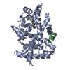
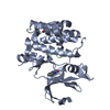
- Assembly
| Deposited unit | 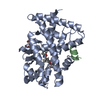
| ||||||||
|---|---|---|---|---|---|---|---|---|---|
| 1 |
| ||||||||
| Unit cell |
|
-Components
| #1: Protein | Mass: 30856.053 Da / Num. of mol.: 1 Source method: isolated from a genetically manipulated source Source: (gene. exp.) Homo sapiens (human) / Plasmid: pET28a / Production host:  |
|---|---|
| #2: Protein/peptide | Mass: 1848.177 Da / Num. of mol.: 1 / Source method: obtained synthetically / Source: (synth.) Homo sapiens (human) / References: UniProt: Q15788, histone acetyltransferase |
| #3: Chemical | ChemComp-P7F / (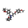  Mass: 490.548 Da / Num. of mol.: 1 / Source method: obtained synthetically / Formula: C28H30N2O6 / Feature type: SUBJECT OF INVESTIGATION Mass: 490.548 Da / Num. of mol.: 1 / Source method: obtained synthetically / Formula: C28H30N2O6 / Feature type: SUBJECT OF INVESTIGATION |
| #4: Chemical | ChemComp-GOL /   Mass: 92.094 Da / Num. of mol.: 1 / Source method: obtained synthetically / Formula: C3H8O3 Mass: 92.094 Da / Num. of mol.: 1 / Source method: obtained synthetically / Formula: C3H8O3 |
| #5: Water | ChemComp-HOH /  Mass: 18.015 Da / Num. of mol.: 111 / Source method: isolated from a natural source / Formula: H2O Mass: 18.015 Da / Num. of mol.: 111 / Source method: isolated from a natural source / Formula: H2O |
| Has ligand of interest | Y |
-Experimental details
-Experiment
| Experiment | Method: X-RAY DIFFRACTION / Number of used crystals: 1 |
|---|
- Sample preparation
| Crystal | Density Matthews: 2.14 Å3/Da / Density % sol: 42.45 % |
|---|---|
| Crystal grow | Temperature: 277 K / Method: vapor diffusion / Details: 0.1M Tris (pH 8.0), 25%(w/v) PEG3350 |
-Data collection
| Diffraction | Mean temperature: 100 K / Serial crystal experiment: N | |||||||||||||||||||||||||||
|---|---|---|---|---|---|---|---|---|---|---|---|---|---|---|---|---|---|---|---|---|---|---|---|---|---|---|---|---|
| Diffraction source | Source: SYNCHROTRON / Site: Photon Factory / Beamline: BL-17A / Wavelength: 1 Å | |||||||||||||||||||||||||||
| Detector | Type: DECTRIS EIGER X 16M / Detector: PIXEL / Date: Mar 7, 2020 / Details: Mirrors | |||||||||||||||||||||||||||
| Radiation | Monochromator: Si(111) / Protocol: SINGLE WAVELENGTH / Monochromatic (M) / Laue (L): M / Scattering type: x-ray | |||||||||||||||||||||||||||
| Radiation wavelength | Wavelength: 1 Å / Relative weight: 1 | |||||||||||||||||||||||||||
| Reflection | Resolution: 1.52→43 Å / Num. obs: 42160 / % possible obs: 99.3 % / Redundancy: 3.4 % / Biso Wilson estimate: 17.89 Å2 / CC1/2: 0.999 / Rmerge(I) obs: 0.037 / Rpim(I) all: 0.023 / Rrim(I) all: 0.043 / Net I/σ(I): 16 | |||||||||||||||||||||||||||
| Reflection shell | Diffraction-ID: 1
|
-Phasing
| Phasing | Method: molecular replacement | |||||||||
|---|---|---|---|---|---|---|---|---|---|---|
| Phasing MR |
|
- Processing
| Software |
| ||||||||||||||||||||||||||||||||||||||||||||||||||||||||||||||||||||||||||||||||||||||||||||||||||||||||||||||||||||||||||||||||||||||||||||||||||||||||||||||||||||||||||||||
|---|---|---|---|---|---|---|---|---|---|---|---|---|---|---|---|---|---|---|---|---|---|---|---|---|---|---|---|---|---|---|---|---|---|---|---|---|---|---|---|---|---|---|---|---|---|---|---|---|---|---|---|---|---|---|---|---|---|---|---|---|---|---|---|---|---|---|---|---|---|---|---|---|---|---|---|---|---|---|---|---|---|---|---|---|---|---|---|---|---|---|---|---|---|---|---|---|---|---|---|---|---|---|---|---|---|---|---|---|---|---|---|---|---|---|---|---|---|---|---|---|---|---|---|---|---|---|---|---|---|---|---|---|---|---|---|---|---|---|---|---|---|---|---|---|---|---|---|---|---|---|---|---|---|---|---|---|---|---|---|---|---|---|---|---|---|---|---|---|---|---|---|---|---|---|---|
| Refinement | Method to determine structure: MOLECULAR REPLACEMENT Starting model: 3SP6 Resolution: 1.52→32.847 Å / SU ML: 0.14 / Cross valid method: FREE R-VALUE / σ(F): 1.91 / Phase error: 21.61 Details: SF FILE CONTAINS FRIEDEL PAIRS UNDER I/F_MINUS AND I/F_PLUS COLUMNS.
| ||||||||||||||||||||||||||||||||||||||||||||||||||||||||||||||||||||||||||||||||||||||||||||||||||||||||||||||||||||||||||||||||||||||||||||||||||||||||||||||||||||||||||||||
| Solvent computation | Shrinkage radii: 0.9 Å / VDW probe radii: 1.11 Å | ||||||||||||||||||||||||||||||||||||||||||||||||||||||||||||||||||||||||||||||||||||||||||||||||||||||||||||||||||||||||||||||||||||||||||||||||||||||||||||||||||||||||||||||
| Displacement parameters | Biso max: 72.63 Å2 / Biso mean: 23.1362 Å2 / Biso min: 10.67 Å2 | ||||||||||||||||||||||||||||||||||||||||||||||||||||||||||||||||||||||||||||||||||||||||||||||||||||||||||||||||||||||||||||||||||||||||||||||||||||||||||||||||||||||||||||||
| Refinement step | Cycle: final / Resolution: 1.52→32.847 Å
| ||||||||||||||||||||||||||||||||||||||||||||||||||||||||||||||||||||||||||||||||||||||||||||||||||||||||||||||||||||||||||||||||||||||||||||||||||||||||||||||||||||||||||||||
| Refine LS restraints |
| ||||||||||||||||||||||||||||||||||||||||||||||||||||||||||||||||||||||||||||||||||||||||||||||||||||||||||||||||||||||||||||||||||||||||||||||||||||||||||||||||||||||||||||||
| LS refinement shell | Refine-ID: X-RAY DIFFRACTION / Rfactor Rfree error: 0
|
