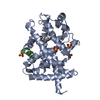
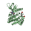
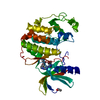
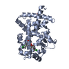
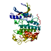
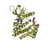
Entry Database : PDB / ID : 3sp6Title Structural basis for iloprost as a dual PPARalpha/delta agonist Peroxisome proliferator-activated receptor alpha Peroxisome proliferator-activated receptor gamma coactivator 1-beta Keywords / / / / Function / homology Function Domain/homology Component
/ / / / / / / / / / / / / / / / / / / / / / / / / / / / / / / / / / / / / / / / / / / / / / / / / / / / / / / / / / / / / / / / / / / / / / / / / / / / / / / / / / / / / / / / / / / / / / / / / / / / / / / / / / / / / / / / / / / / / / / / / / / / / / / / / / / / / / / / / / / / / / / / / Biological species Homo sapiens (human)Method / / / Resolution : 2.21 Å Authors Rong, H. / Li, Y. Journal : J.Biol.Chem. / Year : 2011Title : Structural basis for iloprost as a dual peroxisome proliferator-activated receptor alpha/delta agonist.Authors : Jin, L. / Lin, S. / Rong, H. / Zheng, S. / Jin, S. / Wang, R. / Li, Y. History Deposition Jul 1, 2011 Deposition site / Processing site Revision 1.0 Jul 20, 2011 Provider / Type Revision 1.1 Jun 20, 2012 Group Revision 1.2 Apr 16, 2014 Group Revision 1.3 Feb 28, 2024 Group / Database references / Derived calculationsCategory chem_comp_atom / chem_comp_bond ... chem_comp_atom / chem_comp_bond / database_2 / struct_ref_seq_dif / struct_site Item _database_2.pdbx_DOI / _database_2.pdbx_database_accession ... _database_2.pdbx_DOI / _database_2.pdbx_database_accession / _struct_ref_seq_dif.details / _struct_site.pdbx_auth_asym_id / _struct_site.pdbx_auth_comp_id / _struct_site.pdbx_auth_seq_id
Show all Show less
 Movie
Movie Controller


 Open data
 Basic information
 Components
 Keywords
 Function and homology information
 Homo sapiens (human)
 X-RAY DIFFRACTION /
  Authors
 Citation
 Structure visualization
 Downloads & links
 Other downloads
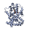


 PDBj
PDBj


 Assembly
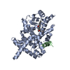


 Mass: 360.487 Da / Num. of mol.: 1 / Source method: obtained synthetically / Formula: C22H32O4
 Mass: 18.015 Da / Num. of mol.: 88 / Source method: isolated from a natural source / Formula: H2O
 Sample preparation
 / Beamline: 21-ID-F / Wavelength: 0.97857 Å
 Processing