Movie
Movie Controller

[English] 日本語
 Yorodumi
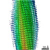
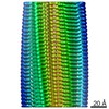
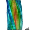
- PDB-7m64: Cryo-EM structure of human islet amyloid polypeptide (hIAPP, or a... -
+ Open data

- Basic information
| Entry | Database: PDB / ID: 7m64 | ||||||
|---|---|---|---|---|---|---|---|
| Title | Cryo-EM structure of human islet amyloid polypeptide (hIAPP, or amylin) fibrils seeded by patient extracted fibrils, polymorph 3 | ||||||
 Components Components | Islet amyloid polypeptide | ||||||
 Keywords Keywords | PROTEIN FIBRIL / hIAPP / type II diabetes / amyloid | ||||||
| Function / homology |  Function and homology information Function and homology informationamylin receptor 3 signaling pathway / amylin receptor 2 signaling pathway / amylin receptor 1 signaling pathway / amylin receptor signaling pathway / Calcitonin-like ligand receptors / negative regulation of amyloid fibril formation / negative regulation of bone resorption / negative regulation of osteoclast differentiation / eating behavior / Regulation of gene expression in beta cells ...amylin receptor 3 signaling pathway / amylin receptor 2 signaling pathway / amylin receptor 1 signaling pathway / amylin receptor signaling pathway / Calcitonin-like ligand receptors / negative regulation of amyloid fibril formation / negative regulation of bone resorption / negative regulation of osteoclast differentiation / eating behavior / Regulation of gene expression in beta cells / positive regulation of cAMP/PKA signal transduction / bone resorption / negative regulation of protein-containing complex assembly / positive regulation of calcium-mediated signaling / osteoclast differentiation / sensory perception of pain / hormone activity / cell-cell signaling / amyloid-beta binding / G alpha (s) signalling events / positive regulation of MAPK cascade / positive regulation of apoptotic process / Amyloid fiber formation / receptor ligand activity / signaling receptor binding / neuronal cell body / apoptotic process / lipid binding / signal transduction / : / extracellular region / identical protein binding Similarity search - Function | ||||||
| Biological species |  Homo sapiens (human) Homo sapiens (human) | ||||||
| Method | ELECTRON MICROSCOPY / helical reconstruction / cryo EM / Resolution: 4 Å | ||||||
 Authors Authors | Cao, Q. / Boyer, D.R. / Sawaya, M.R. / Eisenberg, D.S. | ||||||
| Funding support |  United States, 1items United States, 1items
| ||||||
 Citation Citation | Journal: Nat Struct Mol Biol / Year: 2021 Title: Cryo-EM structures of hIAPP fibrils seeded by patient-extracted fibrils reveal new polymorphs and conserved fibril cores. Authors: Qin Cao / David R Boyer / Michael R Sawaya / Romany Abskharon / Lorena Saelices / Binh A Nguyen / Jiahui Lu / Kevin A Murray / Fouad Kandeel / David S Eisenberg /  Abstract: Amyloidosis of human islet amyloid polypeptide (hIAPP) is a pathological hallmark of type II diabetes (T2D), an epidemic afflicting nearly 10% of the world's population. To visualize disease-relevant ...Amyloidosis of human islet amyloid polypeptide (hIAPP) is a pathological hallmark of type II diabetes (T2D), an epidemic afflicting nearly 10% of the world's population. To visualize disease-relevant hIAPP fibrils, we extracted amyloid fibrils from islet cells of a T2D donor and amplified their quantity by seeding synthetic hIAPP. Cryo-EM studies revealed four fibril polymorphic atomic structures. Their resemblance to four unseeded hIAPP fibrils varies from nearly identical (TW3) to non-existent (TW2). The diverse repertoire of hIAPP polymorphs appears to arise from three distinct protofilament cores entwined in different combinations. The structural distinctiveness of TW1, TW2 and TW4 suggests they may be faithful replications of the pathogenic seeds. If so, the structures determined here provide the most direct view yet of hIAPP amyloid fibrils formed during T2D. | ||||||
| History |
|
- Structure visualization
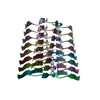
| Movie |
Movie viewer |
|---|---|
| Structure viewer | Molecule: MolmilJmol/JSmol |
- Downloads & links
-Download
| PDBx/mmCIF format | 7m64.cif.gz | 50.1 KB | Display | PDBx/mmCIF format |
|---|---|---|---|---|
| PDB format | pdb7m64.ent.gz | 36.4 KB | Display | PDB format |
| PDBx/mmJSON format | 7m64.json.gz | Tree view | PDBx/mmJSON format | |
| Others |  Other downloads Other downloads |
-Validation report
| Arichive directory | https://data.pdbj.org/pub/pdb/validation_reports/m6/7m64ftp://data.pdbj.org/pub/pdb/validation_reports/m6/7m64 | HTTPS FTP |
|---|
-Related structure data
| Related structure data | 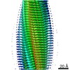 23688MC 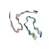 7m61C 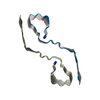 7m62C  7m65C M: map data used to model this data C: citing same article ( |
|---|---|
| Similar structure data | |
| EM raw data | EMPIAR-10868 (Title: Cryo electron microscopy of hIAPP fibrils seeded by patient-extracted fibrils Data size: 9.9 TB Data #1: Unaligned K3 movies of hIAPP fibrils seeded by disease fibrils [micrographs - multiframe]) |

-Links
 PDBj
PDBj


- Assembly
| Deposited unit | 
|
|---|---|
| 1 |
|
| Symmetry | Helical symmetry: (Circular symmetry: 1 / Dyad axis: no / N subunits divisor: 1 / Num. of operations: 10 / Rise per n subunits: 2.4 Å / Rotation per n subunits: 178.33 °) |
-Components
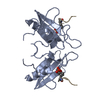
| #1: Protein/peptide | Mass: 3907.312 Da / Num. of mol.: 10 / Fragment: C-terminal amidated peptide (UNP residues 34-70) / Source method: obtained synthetically / Source: (synth.) Homo sapiens (human) / References: UniProt: P10997Has ligand of interest | N | Has protein modification | Y | |
|---|
-Experimental details
-Experiment
| Experiment | Method: ELECTRON MICROSCOPY |
|---|---|
| EM experiment | Aggregation state: FILAMENT / 3D reconstruction method: helical reconstruction |
- Sample preparation
| Component | Name: hIAPP fibril / Type: COMPLEX Details: Synthetic hIAPP seeded by hIAPP fibrils extracted from a patient with Type II Diabetes Entity ID: all / Source: NATURAL |
|---|---|
| Molecular weight | Units: KILODALTONS/NANOMETER / Experimental value: NO |
| Source (natural) | Organism: Homo sapiens (human) |
| Buffer solution | pH: 7.4 |
| Specimen | Embedding applied: NO / Shadowing applied: NO / Staining applied: NO / Vitrification applied: YES |
| Specimen support | Grid material: COPPER / Grid type: Quantifoil R1.2/1.3 |
| Vitrification | Instrument: FEI VITROBOT MARK IV / Cryogen name: ETHANE / Humidity: 100 % / Chamber temperature: 298 K |
- Electron microscopy imaging
| Experimental equipment |  Model: Titan Krios / Image courtesy: FEI Company |
|---|---|
| Microscopy | Model: FEI TITAN KRIOS |
| Electron gun | Electron source:  FIELD EMISSION GUN / Accelerating voltage: 300 kV / Illumination mode: FLOOD BEAM FIELD EMISSION GUN / Accelerating voltage: 300 kV / Illumination mode: FLOOD BEAM |
| Electron lens | Mode: BRIGHT FIELD |
| Image recording | Electron dose: 45 e/Å2 / Detector mode: SUPER-RESOLUTION / Film or detector model: GATAN K3 (6k x 4k) / Num. of grids imaged: 3 |
- Processing
| EM software |
| ||||||||||||||||||||||||||||
|---|---|---|---|---|---|---|---|---|---|---|---|---|---|---|---|---|---|---|---|---|---|---|---|---|---|---|---|---|---|
| CTF correction | Type: PHASE FLIPPING AND AMPLITUDE CORRECTION | ||||||||||||||||||||||||||||
| Helical symmerty | Angular rotation/subunit: 178.33 ° / Axial rise/subunit: 2.4 Å / Axial symmetry: C1 | ||||||||||||||||||||||||||||
| 3D reconstruction | Resolution: 4 Å / Resolution method: FSC 0.143 CUT-OFF / Num. of particles: 17853 / Symmetry type: HELICAL | ||||||||||||||||||||||||||||
| Atomic model building | B value: 83 / Protocol: AB INITIO MODEL / Space: REAL | ||||||||||||||||||||||||||||
| Atomic model building | PDB-ID: 6Y1A Pdb chain-ID: A / Accession code: 6Y1A / Pdb chain residue range: 13-37 / Source name: PDB / Type: experimental model |