 Movie
Movie Controller

[English] 日本語
 Yorodumi
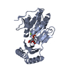
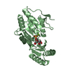
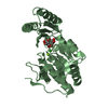
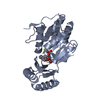
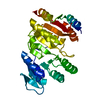
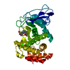
- PDB-6upb: Structure of apo trehalose-6-phosphate phosphatase from Salmonell... -
+ Open data

- Basic information
| Entry | Database: PDB / ID: 6upb | ||||||
|---|---|---|---|---|---|---|---|
| Title | Structure of apo trehalose-6-phosphate phosphatase from Salmonella typhimurium | ||||||
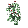
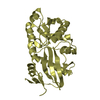
 Components Components | Trehalose-phosphate phosphatase | ||||||
 Keywords Keywords | HYDROLASE / HAD Superfamily / Rossmann fold / sugar binding | ||||||
| Function / homology |  Function and homology information Function and homology informationtrehalose-phosphatase / trehalose-phosphatase activity / trehalose biosynthetic process / magnesium ion binding Similarity search - Function | ||||||
| Biological species |  Salmonella typhimurium (bacteria) Salmonella typhimurium (bacteria) | ||||||
| Method |  X-RAY DIFFRACTION / SYNCHROTRON / MAD / Resolution: 1.89 Å X-RAY DIFFRACTION / SYNCHROTRON / MAD / Resolution: 1.89 Å | ||||||
 Authors Authors | Harvey, C.M. / Allen, K.N. | ||||||
| Funding support |  United States, 1items United States, 1items
| ||||||
 Citation Citation | Journal: Biochemistry / Year: 2020 Title: Structural Analysis of Binding Determinants ofSalmonella typhimuriumTrehalose-6-phosphate Phosphatase Using Ground-State Complexes. Authors: Harvey, C.M. / O'Toole, K.H. / Liu, C. / Mariano, P. / Dunaway-Mariano, D. / Allen, K.N. | ||||||
| History |
|
- Structure visualization
| Structure viewer | Molecule: MolmilJmol/JSmol |
|---|
- Downloads & links
-Download
| PDBx/mmCIF format | 6upb.cif.gz | 205 KB | Display | PDBx/mmCIF format |
|---|---|---|---|---|
| PDB format | pdb6upb.ent.gz | 163.6 KB | Display | PDB format |
| PDBx/mmJSON format | 6upb.json.gz | Tree view | PDBx/mmJSON format | |
| Others |  Other downloads Other downloads |
-Validation report
| Arichive directory | https://data.pdbj.org/pub/pdb/validation_reports/up/6upbftp://data.pdbj.org/pub/pdb/validation_reports/up/6upb | HTTPS FTP |
|---|
-Related structure data


-Links
 PDBj
PDBj- Assembly
| Deposited unit | 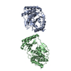
| ||||||||
|---|---|---|---|---|---|---|---|---|---|
| 1 | 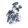
| ||||||||
| 2 | 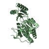
| ||||||||
| Unit cell |
|
-Components
| #1: Protein | Mass: 29296.418 Da / Num. of mol.: 2 Source method: isolated from a genetically manipulated source Source: (gene. exp.) Salmonella typhimurium (strain SL1344) (bacteria)Strain: SL1344 / Gene: otsB, SL1344_1863 / Plasmid: PET15bTEV / Production host: #2: Water | ChemComp-HOH / |  Mass: 18.015 Da / Num. of mol.: 440 / Source method: isolated from a natural source / Formula: H2O Mass: 18.015 Da / Num. of mol.: 440 / Source method: isolated from a natural source / Formula: H2O |
|---|
-Experimental details
-Experiment
| Experiment | Method: X-RAY DIFFRACTION / Number of used crystals: 1 |
|---|
- Sample preparation
| Crystal | Density Matthews: 2.35 Å3/Da / Density % sol: 47.76 % / Description: rod |
|---|---|
| Crystal grow | Temperature: 290 K / Method: vapor diffusion, hanging drop Details: Drop contained: 1.5 uL 40 mg/mL protein, 2 uL reservoir solution (15% PEG 3350, 350 mM lithium citrate), 2 uL water, and 0.5 uL 1:10^5 dilution of seed stock prepared according to Seed Bead ...Details: Drop contained: 1.5 uL 40 mg/mL protein, 2 uL reservoir solution (15% PEG 3350, 350 mM lithium citrate), 2 uL water, and 0.5 uL 1:10^5 dilution of seed stock prepared according to Seed Bead protocol. Crystals were cryoprotected in a solution containing 15% PEG 3350, 350mM lithium citrate, and 10% ethylene glycol |
-Data collection
| Diffraction | Mean temperature: 100 K / Serial crystal experiment: N |
|---|---|
| Diffraction source | Source: SYNCHROTRON / Site: APS / Beamline: 24-ID-E / Wavelength: 0.9791 Å |
| Detector | Type: DECTRIS PILATUS 6M-F / Detector: PIXEL / Date: Feb 17, 2016 |
| Radiation | Protocol: SINGLE WAVELENGTH / Monochromatic (M) / Laue (L): M / Scattering type: x-ray |
| Radiation wavelength | Wavelength: 0.9791 Å / Relative weight: 1 |
| Reflection | Resolution: 1.89→104.4 Å / Num. obs: 39938 / % possible obs: 99.6 % / Redundancy: 8.4 % / Biso Wilson estimate: 24.52 Å2 / CC1/2: 0.998 / Rmerge(I) obs: 0.109 / Net I/σ(I): 14.9 |
| Reflection shell | Resolution: 1.89→1.958 Å / Redundancy: 8.1 % / Rmerge(I) obs: 0.6532 / Mean I/σ(I) obs: 3.36 / Num. unique obs: 3940 / CC1/2: 0.846 / % possible all: 99.6 |
- Processing
| Software |
| ||||||||||||||||||||||||||||||||||||||||||||||||||||||||||||||||||||||||||||||
|---|---|---|---|---|---|---|---|---|---|---|---|---|---|---|---|---|---|---|---|---|---|---|---|---|---|---|---|---|---|---|---|---|---|---|---|---|---|---|---|---|---|---|---|---|---|---|---|---|---|---|---|---|---|---|---|---|---|---|---|---|---|---|---|---|---|---|---|---|---|---|---|---|---|---|---|---|---|---|---|
| Refinement | Method to determine structure: MAD / Resolution: 1.89→104.4 Å / SU ML: 0.19 / Cross valid method: THROUGHOUT / σ(F): 1.37 / Phase error: 21.95
| ||||||||||||||||||||||||||||||||||||||||||||||||||||||||||||||||||||||||||||||
| Solvent computation | Shrinkage radii: 0.9 Å / VDW probe radii: 1.11 Å | ||||||||||||||||||||||||||||||||||||||||||||||||||||||||||||||||||||||||||||||
| Displacement parameters | Biso max: 90.51 Å2 / Biso mean: 34.3761 Å2 / Biso min: 11.62 Å2 | ||||||||||||||||||||||||||||||||||||||||||||||||||||||||||||||||||||||||||||||
| Refinement step | Cycle: final / Resolution: 1.89→104.4 Å
| ||||||||||||||||||||||||||||||||||||||||||||||||||||||||||||||||||||||||||||||
| LS refinement shell | Refine-ID: X-RAY DIFFRACTION / Rfactor Rfree error: 0
|
