[English] 日本語
Yorodumi
- PDB-2fea: Crystal structure of MtnX phosphatase from Bacillus Subtilis at 2... -

+
Open data


ID or keywords:

Loading...

-
Basic information

Entry
Database: PDB / ID: 2fea
TitleCrystal structure of MtnX phosphatase from Bacillus Subtilis at 2.00 A resolution
Components2-hydroxy-3-keto-5-methylthiopentenyl-1-phosphate phosphatase
KeywordsHYDROLASE / 2633731 / Structural Genomics / Joint Center for Structural Genomics / JCSG / Protein Structure Initiative / PSI
Function / homology
Function and homology information


2-hydroxy-3-keto-5-methylthiopentenyl-1-phosphate phosphatase / 2-hydroxy-3-keto-5-methylthiopentenyl-1-phosphate phosphatase activity / L-methionine salvage from S-adenosylmethionine / L-methionine salvage from methylthioadenosine
Similarity search - Function
thrh gene product, domain 2 - #20 / HAD hydrolase, subfamily IA, Pyridoxal phosphate phosphatase-like / 2-hydroxy-3-keto-5-methylthiopentenyl-1-phosphate phosphatase / thrh gene product, domain 2 / haloacid dehalogenase-like hydrolase / HAD superfamily/HAD-like / HAD superfamily / HAD-like superfamily / Alpha-Beta Complex / Rossmann fold ...thrh gene product, domain 2 - #20 / HAD hydrolase, subfamily IA, Pyridoxal phosphate phosphatase-like / 2-hydroxy-3-keto-5-methylthiopentenyl-1-phosphate phosphatase / thrh gene product, domain 2 / haloacid dehalogenase-like hydrolase / HAD superfamily/HAD-like / HAD superfamily / HAD-like superfamily / Alpha-Beta Complex / Rossmann fold / 3-Layer(aba) Sandwich / Alpha Beta
Similarity search - Domain/homology
2-hydroxy-3-keto-5-methylthiopentenyl-1-phosphate phosphatase
Similarity search - Component
Biological speciesBacillus subtilis (bacteria)
MethodX-RAY DIFFRACTION / SYNCHROTRON / MAD / Resolution: 2 Å
AuthorsJoint Center for Structural Genomics (JCSG)
CitationJournal: Proteins / Year: 2007
Title: Crystal structure of MtnX phosphatase from Bacillus subtilis at 2.0 A resolution provides a structural basis for bipartite phosphomonoester hydrolysis of 2-hydroxy-3-keto-5-methylthiopentenyl-1-phosphate.
Authors: Xu, Q. / Saikatendu, K.S. / Krishna, S.S. / McMullan, D. / Abdubek, P. / Agarwalla, S. / Ambing, E. / Astakhova, T. / Axelrod, H.L. / Carlton, D. / Chiu, H.J. / Clayton, T. / DiDonato, M. / ...Authors: Xu, Q. / Saikatendu, K.S. / Krishna, S.S. / McMullan, D. / Abdubek, P. / Agarwalla, S. / Ambing, E. / Astakhova, T. / Axelrod, H.L. / Carlton, D. / Chiu, H.J. / Clayton, T. / DiDonato, M. / Duan, L. / Elsliger, M.A. / Feuerhelm, J. / Grzechnik, S.K. / Hale, J. / Hampton, E. / Han, G.W. / Haugen, J. / Jaroszewski, L. / Jin, K.K. / Klock, H.E. / Knuth, M.W. / Koesema, E. / Miller, M.D. / Morse, A.T. / Nigoghossian, E. / Okach, L. / Oommachen, S. / Paulsen, J. / Reyes, R. / Rife, C.L. / Schwarzenbacher, R. / van den Bedem, H. / White, A. / Wolf, G. / Hodgson, K.O. / Wooley, J. / Deacon, A.M. / Godzik, A. / Lesley, S.A. / Wilson, I.A.
History
DepositionDec 15, 2005Deposition site: RCSB / Processing site: RCSB
Revision 1.0Dec 27, 2005Provider: repository / Type: Initial release
Revision 1.1May 1, 2008Group: Version format compliance
Revision 1.2Jul 13, 2011Group: Advisory / Version format compliance
Revision 1.3Jan 25, 2023Group: Database references / Derived calculations
Category: database_2 / pdbx_struct_conn_angle ...database_2 / pdbx_struct_conn_angle / struct_conn / struct_conn_type / struct_ref_seq_dif / struct_site
Item: _database_2.pdbx_DOI / _database_2.pdbx_database_accession ..._database_2.pdbx_DOI / _database_2.pdbx_database_accession / _pdbx_struct_conn_angle.ptnr1_auth_asym_id / _pdbx_struct_conn_angle.ptnr1_auth_comp_id / _pdbx_struct_conn_angle.ptnr1_auth_seq_id / _pdbx_struct_conn_angle.ptnr1_label_asym_id / _pdbx_struct_conn_angle.ptnr1_label_atom_id / _pdbx_struct_conn_angle.ptnr1_label_comp_id / _pdbx_struct_conn_angle.ptnr1_label_seq_id / _pdbx_struct_conn_angle.ptnr2_auth_asym_id / _pdbx_struct_conn_angle.ptnr2_auth_comp_id / _pdbx_struct_conn_angle.ptnr2_auth_seq_id / _pdbx_struct_conn_angle.ptnr2_label_asym_id / _pdbx_struct_conn_angle.ptnr2_label_atom_id / _pdbx_struct_conn_angle.ptnr2_label_comp_id / _pdbx_struct_conn_angle.ptnr3_auth_asym_id / _pdbx_struct_conn_angle.ptnr3_auth_comp_id / _pdbx_struct_conn_angle.ptnr3_auth_seq_id / _pdbx_struct_conn_angle.ptnr3_label_asym_id / _pdbx_struct_conn_angle.ptnr3_label_atom_id / _pdbx_struct_conn_angle.ptnr3_label_comp_id / _pdbx_struct_conn_angle.ptnr3_label_seq_id / _pdbx_struct_conn_angle.value / _struct_conn.conn_type_id / _struct_conn.id / _struct_conn.pdbx_dist_value / _struct_conn.pdbx_leaving_atom_flag / _struct_conn.ptnr1_auth_asym_id / _struct_conn.ptnr1_auth_comp_id / _struct_conn.ptnr1_auth_seq_id / _struct_conn.ptnr1_label_asym_id / _struct_conn.ptnr1_label_atom_id / _struct_conn.ptnr1_label_comp_id / _struct_conn.ptnr1_label_seq_id / _struct_conn.ptnr2_auth_asym_id / _struct_conn.ptnr2_auth_comp_id / _struct_conn.ptnr2_auth_seq_id / _struct_conn.ptnr2_label_asym_id / _struct_conn.ptnr2_label_atom_id / _struct_conn.ptnr2_label_comp_id / _struct_conn.ptnr2_label_seq_id / _struct_conn_type.id / _struct_ref_seq_dif.details / _struct_site.pdbx_auth_asym_id / _struct_site.pdbx_auth_comp_id / _struct_site.pdbx_auth_seq_id
Remark 999SEQUENCE THE CONSTRUCT WAS EXPRESSED WITH A PURIFICATION TAG MGSDKIHHHHHHENLYFQG. THE TAG WAS ...SEQUENCE THE CONSTRUCT WAS EXPRESSED WITH A PURIFICATION TAG MGSDKIHHHHHHENLYFQG. THE TAG WAS REMOVED WITH TEV PROTEASE LEAVING ONLY A GLYCINE (0) FOLLOWED BY THE TARGET SEQUENCE.

-
Structure visualization

Structure viewerMolecule:
MolmilJmol/JSmol

Downloads & links

-
Assembly

Deposited unit
A: 2-hydroxy-3-keto-5-methylthiopentenyl-1-phosphate phosphatase
B: 2-hydroxy-3-keto-5-methylthiopentenyl-1-phosphate phosphatase
hetero molecules


Theoretical massNumber of molelcules
Total (without water)55,63216
Polymers54,8322
Non-polymers80014
Water6,521362
1
A: 2-hydroxy-3-keto-5-methylthiopentenyl-1-phosphate phosphatase
hetero molecules


Theoretical massNumber of molelcules
Total (without water)27,7547
Polymers27,4161
Non-polymers3386
Water181
TypeNameSymmetry operationNumber
identity operation1_555x,y,z1
2
B: 2-hydroxy-3-keto-5-methylthiopentenyl-1-phosphate phosphatase
hetero molecules


Theoretical massNumber of molelcules
Total (without water)27,8789
Polymers27,4161
Non-polymers4628
Water181
TypeNameSymmetry operationNumber
identity operation1_555x,y,z1
Unit cell
Length a, b, c (Å)59.013, 43.104, 109.553
Angle α, β, γ (deg.)90.000, 98.910, 90.000
Int Tables number4
Space group name H-MP1211
Noncrystallographic symmetry (NCS)NCS domain:
IDEns-IDDetails
11A
21B

NCS domain segments:

Component-ID: 1 / Ens-ID: 1 / Beg label comp-ID: THR / End label comp-ID: ASN / Refine code: 4 / Auth seq-ID: 3 - 226 / Label seq-ID: 4 - 227

Dom-IDAuth asym-IDLabel asym-ID
1AA
2BB

-
Components

#1: Protein 2-hydroxy-3-keto-5-methylthiopentenyl-1-phosphate phosphatase / HK-MTPenyl-1-P phosphatase


Mass: 27416.141 Da / Num. of mol.: 2
Source method: isolated from a genetically manipulated source
Source: (gene. exp.) Bacillus subtilis (bacteria) / Gene: mtnX / Production host: Escherichia coli (E. coli)
References: UniProt: O31667, Hydrolases; Acting on ester bonds; Phosphoric-monoester hydrolases
#2: Chemical ChemComp-MG / MAGNESIUM ION


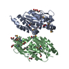
Mass: 24.305 Da / Num. of mol.: 2 / Source method: obtained synthetically / Formula: Mg
#3: Chemical ChemComp-ZN / ZINC ION


Mass: 65.409 Da / Num. of mol.: 2 / Source method: obtained synthetically / Formula: Zn
#4: Chemical
ChemComp-EDO / 1,2-ETHANEDIOL / ETHYLENE GLYCOL


Mass: 62.068 Da / Num. of mol.: 10 / Source method: obtained synthetically / Formula: C2H6O2
#5: Water ChemComp-HOH / water


Mass: 18.015 Da / Num. of mol.: 362 / Source method: isolated from a natural source / Formula: H2O

-
Experimental details

-
Experiment

ExperimentMethod: X-RAY DIFFRACTION / Number of used crystals: 1

-
Sample preparation

CrystalDensity Matthews: 2.42 Å3/Da / Density % sol: 48.8 %
Crystal growTemperature: 277 K / Method: vapor diffusion, sitting drop, nanodrop / pH: 8.5
Details: 0.2M NaOAc, 30.0% PEG-4000, 0.1M TRIS, pH 8.5, VAPOR DIFFUSION,SITTING DROP,NANODROP, temperature 277K

-
Data collection

DiffractionMean temperature: 100 K
Diffraction sourceSource: SYNCHROTRON / Site: ALS / Beamline: 8.3.1 / Wavelength: 1.019867, 0.979741
DetectorType: ADSC QUANTUM 210 / Detector: CCD / Date: Oct 12, 2005
RadiationMonochromator: Double Crystal Si(111) / Protocol: MAD / Monochromatic (M) / Laue (L): M / Scattering type: x-ray
Radiation wavelength
IDWavelength (Å)Relative weight
11.0198671
20.9797411
ReflectionResolution: 2→22.95 Å / Num. obs: 36411 / % possible obs: 98 % / Redundancy: 3.9 % / Rmerge(I) obs: 0.061 / Rsym value: 0.061 / Net I/σ(I): 8.1
Reflection shell

Diffraction-ID: 1

Resolution (Å)% possible obs (%)Redundancy (%)Rmerge(I) obsMean I/σ(I) obsNum. measured obsRsym value% possible all
2-2.05973.80.4181.826580.41897
2.05-2.1196.93.90.3981.125600.398
2.11-2.1797.340.2972.525260.297
2.17-2.2497.540.233.224160.23
2.24-2.3197.540.221.423660.22
2.31-2.3997.740.164.623190.16
2.39-2.4897.840.145.222260.14
2.48-2.5897.940.1166.321630.116
2.58-2.798.23.90.1055.320790.105
2.7-2.8398.240.0749.519630.074
2.83-2.9898.540.06510.518800.065
2.98-3.1698.540.05412.618000.054
3.16-3.3898.940.04614.216840.046
3.38-3.6598.93.90.04313.316000.043
3.65-498.93.80.04111.114400.041
4-4.47993.90.03517.513240.035
4.47-5.1699.23.90.03517.511880.035
5.16-6.3299.43.80.03716.29980.037
6.32-8.9499.33.70.035167970.035
8.94-22.95933.30.02921.34240.029

-
Phasing

PhasingMethod: MAD

-
Processing

Software
NameVersionClassificationNB
REFMAC5.2.0005refinement
SCALAdata scaling
PDB_EXTRACT1.601data extraction
MOSFLMdata reduction
CCP4(SCALA)data scaling
SOLVEphasing
RefinementMethod to determine structure: MAD / Resolution: 2→22.95 Å / Cor.coef. Fo:Fc: 0.969 / Cor.coef. Fo:Fc free: 0.942 / SU B: 8.124 / SU ML: 0.116 / TLS residual ADP flag: LIKELY RESIDUAL / Cross valid method: THROUGHOUT / ESU R: 0.154 / ESU R Free: 0.152
Stereochemistry target values: MAXIMUM LIKELIHOOD WITH PHASES
Details: 1. HYDROGENS HAVE BEEN ADDED IN THE RIDING POSITIONS. 2. A MET-INHIBITION PROTOCOL WAS USED FOR SELENOMETHIONINE INCORPORATION DURING PROTEIN EXPRESSION. THE OCCUPANCY OF THE SE ATOMS IN THE ...Details: 1. HYDROGENS HAVE BEEN ADDED IN THE RIDING POSITIONS. 2. A MET-INHIBITION PROTOCOL WAS USED FOR SELENOMETHIONINE INCORPORATION DURING PROTEIN EXPRESSION. THE OCCUPANCY OF THE SE ATOMS IN THE MSE RESIDUES WAS REDUCED TO 0.7 TO ACCOUNT FOR THE REDUCED SCATTERING POWER DUE TO PARTIAL S-MET INCORPORATION. 3. METAL SITES: TWO METAL SITES WERE IDENTIFIED IN EACH MONOMER. MAGNESIUM, LOCATED AT THE ACTIVE SITE, WAS MODELLED BASED ON STRUCTURAL HOMOLOGS; THE OTHER METAL SITE IS COORDINATED BY FOUR CYSTEINES: 139,143,147,150. IT WAS TENTATIVELY ASSIGNED AS ZINC.
RfactorNum. reflection% reflectionSelection details
Rfree0.217 1810 5 %RANDOM
Rwork0.16 ---
all0.163 ---
obs0.16289 34588 97.55 %-
Solvent computationIon probe radii: 0.8 Å / Shrinkage radii: 0.8 Å / VDW probe radii: 1.2 Å / Solvent model: BABINET MODEL WITH MASK
Displacement parametersBiso mean: 35.926 Å2
Baniso -1Baniso -2Baniso -3
1--0.08 Å20 Å20.12 Å2
2--2.15 Å20 Å2
3----2.03 Å2
Refinement stepCycle: LAST / Resolution: 2→22.95 Å
ProteinNucleic acidLigandSolventTotal
Num. atoms3613 0 44 362 4019
Refine LS restraints
Refine-IDTypeDev idealDev ideal targetNumber
X-RAY DIFFRACTIONr_bond_refined_d0.0180.0223770
X-RAY DIFFRACTIONr_bond_other_d0.0020.023352
X-RAY DIFFRACTIONr_angle_refined_deg1.481.9565091
X-RAY DIFFRACTIONr_angle_other_deg0.85737835
X-RAY DIFFRACTIONr_dihedral_angle_1_deg6.0285461
X-RAY DIFFRACTIONr_dihedral_angle_2_deg36.10724.866187
X-RAY DIFFRACTIONr_dihedral_angle_3_deg14.81715653
X-RAY DIFFRACTIONr_dihedral_angle_4_deg13.9171517
X-RAY DIFFRACTIONr_chiral_restr0.0930.2549
X-RAY DIFFRACTIONr_gen_planes_refined0.0060.024174
X-RAY DIFFRACTIONr_gen_planes_other0.0010.02761
X-RAY DIFFRACTIONr_nbd_refined0.2240.2783
X-RAY DIFFRACTIONr_nbd_other0.1820.23456
X-RAY DIFFRACTIONr_nbtor_refined0.1870.21862
X-RAY DIFFRACTIONr_nbtor_other0.0840.22154
X-RAY DIFFRACTIONr_xyhbond_nbd_refined0.1680.2278
X-RAY DIFFRACTIONr_symmetry_vdw_refined0.1120.21
X-RAY DIFFRACTIONr_symmetry_vdw_other0.2520.234
X-RAY DIFFRACTIONr_symmetry_hbond_refined0.160.217
X-RAY DIFFRACTIONr_mcbond_it1.94432322
X-RAY DIFFRACTIONr_mcbond_other0.6563911
X-RAY DIFFRACTIONr_mcangle_it2.85953682
X-RAY DIFFRACTIONr_scbond_it5.52281636
X-RAY DIFFRACTIONr_scangle_it7.524111402
Refine LS restraints NCS

Dom-ID: 1 / Auth asym-ID: A / Ens-ID: 1 / Number: 3354 / Refine-ID: X-RAY DIFFRACTION

TypeRms dev position (Å)Weight position
medium positional0.60.5
medium thermal1.022
LS refinement shellResolution: 2→2.052 Å / Total num. of bins used: 20
RfactorNum. reflection% reflection
Rfree0.288 123 -
Rwork0.207 2532 -
obs--96.76 %
Refinement TLS params.

Method: refined / Refine-ID: X-RAY DIFFRACTION

IDL112)L122)L132)L222)L232)L332)S11 (Å °)S12 (Å °)S13 (Å °)S21 (Å °)S22 (Å °)S23 (Å °)S31 (Å °)S32 (Å °)S33 (Å °)T112)T122)T132)T222)T232)T332)Origin x (Å)Origin y (Å)Origin z (Å)
12.1750.3542-0.68621.38540.06422.48380.0703-0.05420.03010.0257-0.05530.1384-0.0173-0.2762-0.015-0.14060.0281-0.0085-0.1659-0.0085-0.157748.69870.038740.1858
23.3749-1.4302-0.12911.46720.60462.21360.03940.23680.04510.0240.0566-0.1350.21050.304-0.096-0.15190.0123-0.0151-0.1334-0.0019-0.126433.9379-7.27514.3112
Refinement TLS group
IDRefine-IDRefine TLS-IDAuth asym-IDLabel asym-IDAuth seq-IDLabel seq-ID
1X-RAY DIFFRACTION1AA2 - 2273 - 228
2X-RAY DIFFRACTION1AC13011
3X-RAY DIFFRACTION1AD13021
4X-RAY DIFFRACTION2BB3 - 2264 - 227
5X-RAY DIFFRACTION2BE23011
6X-RAY DIFFRACTION2BF23021

+
About Yorodumi

-
News

-
Feb 9, 2022. New format data for meta-information of EMDB entries

New format data for meta-information of EMDB entries

  • Version 3 of the EMDB header file is now the official format.
  • The previous official version 1.9 will be removed from the archive.

Related info.:EMDB header

External links:wwPDB to switch to version 3 of the EMDB data model

-
Aug 12, 2020. Covid-19 info

Covid-19 info

URL: https://pdbjlvh1.pdbj.org/emnavi/covid19.php

New page: Covid-19 featured information page in EM Navigator.

Related info.:Covid-19 info / Mar 5, 2020. Novel coronavirus structure data

+
Mar 5, 2020. Novel coronavirus structure data

Novel coronavirus structure data

Related info.:Yorodumi Speices / Aug 12, 2020. Covid-19 info

External links:COVID-19 featured content - PDBj / Molecule of the Month (242):Coronavirus Proteases

+
Jan 31, 2019. EMDB accession codes are about to change! (news from PDBe EMDB page)

EMDB accession codes are about to change! (news from PDBe EMDB page)

  • The allocation of 4 digits for EMDB accession codes will soon come to an end. Whilst these codes will remain in use, new EMDB accession codes will include an additional digit and will expand incrementally as the available range of codes is exhausted. The current 4-digit format prefixed with “EMD-” (i.e. EMD-XXXX) will advance to a 5-digit format (i.e. EMD-XXXXX), and so on. It is currently estimated that the 4-digit codes will be depleted around Spring 2019, at which point the 5-digit format will come into force.
  • The EM Navigator/Yorodumi systems omit the EMD- prefix.

Related info.:Q: What is EMD? / ID/Accession-code notation in Yorodumi/EM Navigator

External links:EMDB Accession Codes are Changing Soon! / Contact to PDBj

+
Jul 12, 2017. Major update of PDB

Major update of PDB

  • wwPDB released updated PDB data conforming to the new PDBx/mmCIF dictionary.
  • This is a major update changing the version number from 4 to 5, and with Remediation, in which all the entries are updated.
  • In this update, many items about electron microscopy experimental information are reorganized (e.g. em_software).
  • Now, EM Navigator and Yorodumi are based on the updated data.

External links:wwPDB Remediation / Enriched Model Files Conforming to OneDep Data Standards Now Available in the PDB FTP Archive

-
Yorodumi

Thousand views of thousand structures

  • Yorodumi is a browser for structure data from EMDB, PDB, SASBDB, etc.
  • This page is also the successor to EM Navigator detail page, and also detail information page/front-end page for Omokage search.
  • The word "yorodu" (or yorozu) is an old Japanese word meaning "ten thousand". "mi" (miru) is to see.

Related info.:EMDB / PDB / SASBDB / Comparison of 3 databanks / Yorodumi Search / Aug 31, 2016. New EM Navigator & Yorodumi / Yorodumi Papers / Jmol/JSmol / Function and homology information / Changes in new EM Navigator and Yorodumi

Read more