 Movie
Movie Controller

[English] 日本語
 Yorodumi
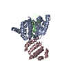
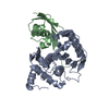
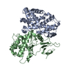
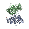
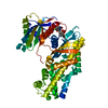
- PDB-2xre: Detection of cobalt in previously unassigned human SENP1 structure -
+ Open data

- Basic information
| Entry | Database: PDB / ID: 2xre | ||||||
|---|---|---|---|---|---|---|---|
| Title | Detection of cobalt in previously unassigned human SENP1 structure | ||||||
 Components Components | SENTRIN-SPECIFIC PROTEASE 1 | ||||||
 Keywords Keywords | HYDROLASE / CYSTEINE PROTEASE | ||||||
| Function / homology |  Function and homology information Function and homology informationregulation of definitive erythrocyte differentiation / SUMO-specific endopeptidase activity / deSUMOylase activity / protein desumoylation / SUMO is proteolytically processed / RHOF GTPase cycle / regulation of postsynapse assembly / protein sumoylation / postsynaptic cytosol / presynaptic cytosol ...regulation of definitive erythrocyte differentiation / SUMO-specific endopeptidase activity / deSUMOylase activity / protein desumoylation / SUMO is proteolytically processed / RHOF GTPase cycle / regulation of postsynapse assembly / protein sumoylation / postsynaptic cytosol / presynaptic cytosol / regulation of mRNA stability / negative regulation of proteasomal ubiquitin-dependent protein catabolic process / nuclear membrane / endopeptidase activity / proteasome-mediated ubiquitin-dependent protein catabolic process / Hydrolases; Acting on peptide bonds (peptidases); Cysteine endopeptidases / focal adhesion / glutamatergic synapse / positive regulation of transcription by RNA polymerase II / nucleoplasm / nucleus Similarity search - Function | ||||||
| Biological species |  HOMO SAPIENS (human) HOMO SAPIENS (human) | ||||||
| Method |  X-RAY DIFFRACTION / SYNCHROTRON / MOLECULAR REPLACEMENT / Resolution: 2.45 Å X-RAY DIFFRACTION / SYNCHROTRON / MOLECULAR REPLACEMENT / Resolution: 2.45 Å | ||||||
 Authors Authors | Rimsa, V. / Eadsforth, T. / Hay, R.T. / Hunter, W.N. | ||||||
 Citation Citation | Journal: Acta Crystallogr.,Sect.F / Year: 2011 Title: The Role of Co2+ in the Crystallization of Human Senp1 and Comments on the Limitations of Automated Refinement Protocols Authors: Rimsa, V. / Eadsforth, T. / Hunter, W.N. | ||||||
| History |
|
- Structure visualization
| Structure viewer | Molecule: MolmilJmol/JSmol |
|---|
- Downloads & links
-Download
| PDBx/mmCIF format | 2xre.cif.gz | 111.9 KB | Display | PDBx/mmCIF format |
|---|---|---|---|---|
| PDB format | pdb2xre.ent.gz | 86.5 KB | Display | PDB format |
| PDBx/mmJSON format | 2xre.json.gz | Tree view | PDBx/mmJSON format | |
| Others |  Other downloads Other downloads |
-Validation report
| Arichive directory | https://data.pdbj.org/pub/pdb/validation_reports/xr/2xreftp://data.pdbj.org/pub/pdb/validation_reports/xr/2xre | HTTPS FTP |
|---|
-Related structure data
| Related structure data |  2xphC  2iycS S: Starting model for refinement C: citing same article ( |
|---|---|
| Similar structure data |


-Links
 PDBj
PDBj
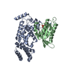
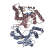
- Assembly
| Deposited unit | 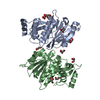
| ||||||||
|---|---|---|---|---|---|---|---|---|---|
| 1 |
| ||||||||
| 2 | 
| ||||||||
| 3 | 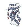
| ||||||||
| 4 | 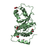
| ||||||||
| Unit cell |
|
-Components
| #1: Protein | Mass: 27378.580 Da / Num. of mol.: 2 / Fragment: CATALYTIC FRAGMENT, RESIDUES 415-644 Source method: isolated from a genetically manipulated source Details: COBALT(II) ION, GLYCEROL / Source: (gene. exp.) HOMO SAPIENS (human) / Cell line: ESCHERICHIA COLI / Plasmid: PHISTEV30A / Production host:  References: UniProt: Q9P0U3, Hydrolases; Acting on peptide bonds (peptidases); Cysteine endopeptidases #2: Chemical | ChemComp-GOL /   Mass: 92.094 Da / Num. of mol.: 10 / Source method: obtained synthetically / Formula: C3H8O3 Mass: 92.094 Da / Num. of mol.: 10 / Source method: obtained synthetically / Formula: C3H8O3#3: Chemical | ChemComp-CO / |   Mass: 58.933 Da / Num. of mol.: 1 / Source method: obtained synthetically / Formula: Co Mass: 58.933 Da / Num. of mol.: 1 / Source method: obtained synthetically / Formula: Co#4: Water | ChemComp-HOH / |  Mass: 18.015 Da / Num. of mol.: 61 / Source method: isolated from a natural source / Formula: H2O Mass: 18.015 Da / Num. of mol.: 61 / Source method: isolated from a natural source / Formula: H2O |
|---|
-Experimental details
-Experiment
| Experiment | Method: X-RAY DIFFRACTION |
|---|
- Sample preparation
| Crystal | Density Matthews: 2.74 Å3/Da / Density % sol: 55.11 % / Description: NONE |
|---|---|
| Crystal grow | Method: vapor diffusion, sitting drop Details: CRYSTALLIZATION WAS PERFORMED USING A SITTING-DROP VAPOUR-DIFFUSION METHOD. CRYSTALS APPEARED FROM EQUAL VOLUMES OF PROTEIN SOLUTION (20 MG/ML IN 20 MM TRIS/HCL, PH 8.0, AND 50 MM NACL) AND ...Details: CRYSTALLIZATION WAS PERFORMED USING A SITTING-DROP VAPOUR-DIFFUSION METHOD. CRYSTALS APPEARED FROM EQUAL VOLUMES OF PROTEIN SOLUTION (20 MG/ML IN 20 MM TRIS/HCL, PH 8.0, AND 50 MM NACL) AND RESERVOIR SOLUTION CONTAINING 100 MM COCL2, 0.1M MES, PH 6.5, AND 1.8 M (NH4)2SO4. |
-Data collection
| Diffraction | Mean temperature: 100 K |
|---|---|
| Diffraction source | Source: SYNCHROTRON / Site: ESRF  / Beamline: ID14-4 / Wavelength: 0.979 / Beamline: ID14-4 / Wavelength: 0.979 |
| Detector | Type: ADSC CCD / Detector: CCD |
| Radiation | Protocol: SINGLE WAVELENGTH / Monochromatic (M) / Laue (L): M / Scattering type: x-ray |
| Radiation wavelength | Wavelength: 0.979 Å / Relative weight: 1 |
| Reflection | Resolution: 2.45→45.6 Å / Num. obs: 21832 / % possible obs: 100 % / Observed criterion σ(I): 0 / Redundancy: 8.8 % / Rmerge(I) obs: 0.1 / Net I/σ(I): 19 |
| Reflection shell | Resolution: 2.45→2.51 Å / Rmerge(I) obs: 0.43 / Mean I/σ(I) obs: 3 / % possible all: 100 |
- Processing
| Software |
| ||||||||||||||||||||||||||||||||||||||||||||||||||||||||||||||||||||||||||||||||||||||||||||||||||||||||||||||||||||||||||||||||||||||||||||||||||||||||||||||||||||||||||||||||||||||
|---|---|---|---|---|---|---|---|---|---|---|---|---|---|---|---|---|---|---|---|---|---|---|---|---|---|---|---|---|---|---|---|---|---|---|---|---|---|---|---|---|---|---|---|---|---|---|---|---|---|---|---|---|---|---|---|---|---|---|---|---|---|---|---|---|---|---|---|---|---|---|---|---|---|---|---|---|---|---|---|---|---|---|---|---|---|---|---|---|---|---|---|---|---|---|---|---|---|---|---|---|---|---|---|---|---|---|---|---|---|---|---|---|---|---|---|---|---|---|---|---|---|---|---|---|---|---|---|---|---|---|---|---|---|---|---|---|---|---|---|---|---|---|---|---|---|---|---|---|---|---|---|---|---|---|---|---|---|---|---|---|---|---|---|---|---|---|---|---|---|---|---|---|---|---|---|---|---|---|---|---|---|---|---|
| Refinement | Method to determine structure: MOLECULAR REPLACEMENT Starting model: PDB ENTRY 2IYC Resolution: 2.45→45.6 Å / Cor.coef. Fo:Fc: 0.927 / Cor.coef. Fo:Fc free: 0.874 / SU B: 11.596 / SU ML: 0.27 / Cross valid method: THROUGHOUT / ESU R: 0.479 / ESU R Free: 0.337 / Stereochemistry target values: MAXIMUM LIKELIHOOD / Details: HYDROGENS HAVE BEEN ADDED IN THE RIDING POSITIONS
| ||||||||||||||||||||||||||||||||||||||||||||||||||||||||||||||||||||||||||||||||||||||||||||||||||||||||||||||||||||||||||||||||||||||||||||||||||||||||||||||||||||||||||||||||||||||
| Solvent computation | Ion probe radii: 0.8 Å / Shrinkage radii: 0.8 Å / VDW probe radii: 1.4 Å / Solvent model: MASK | ||||||||||||||||||||||||||||||||||||||||||||||||||||||||||||||||||||||||||||||||||||||||||||||||||||||||||||||||||||||||||||||||||||||||||||||||||||||||||||||||||||||||||||||||||||||
| Displacement parameters | Biso mean: 51.36 Å2
| ||||||||||||||||||||||||||||||||||||||||||||||||||||||||||||||||||||||||||||||||||||||||||||||||||||||||||||||||||||||||||||||||||||||||||||||||||||||||||||||||||||||||||||||||||||||
| Refinement step | Cycle: LAST / Resolution: 2.45→45.6 Å
| ||||||||||||||||||||||||||||||||||||||||||||||||||||||||||||||||||||||||||||||||||||||||||||||||||||||||||||||||||||||||||||||||||||||||||||||||||||||||||||||||||||||||||||||||||||||
| Refine LS restraints |
|