FHA domain binding / positive regulation of ligase activity / DNA ligase IV complex / DNA double-strand break attachment to nuclear envelope / DNA-dependent protein kinase-DNA ligase 4 complex / immunoglobulin V(D)J recombination / nonhomologous end joining complex / spindle pole body / protein localization to site of double-strand break / cellular response to lithium ion ...FHA domain binding / positive regulation of ligase activity / DNA ligase IV complex / DNA double-strand break attachment to nuclear envelope / DNA-dependent protein kinase-DNA ligase 4 complex / immunoglobulin V(D)J recombination / nonhomologous end joining complex / spindle pole body / protein localization to site of double-strand break / cellular response to lithium ion / 2-LTR circle formation / response to X-ray / SUMOylation of DNA damage response and repair proteins / Nonhomologous End-Joining (NHEJ) / base-excision repair / double-strand break repair via nonhomologous end joining / double-strand break repair / site of double-strand break / protein-macromolecule adaptor activity / enzyme binding / DNA binding / nucleoplasm / identical protein binding / nucleus / cytosol Similarity search - Function
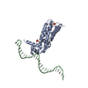
Spindle pole body component 110, C-terminal / Spindle pole body component 110 C-terminal domain / XRCC4, N-terminal domain superfamily / DNA repair protein XRCC4 / : / : / : / XRCC4 N-terminal domain / XRCC4 coiled-coil / XRCC4 C-terminal region ...Spindle pole body component 110, C-terminal / Spindle pole body component 110 C-terminal domain / XRCC4, N-terminal domain superfamily / DNA repair protein XRCC4 / : / : / : / XRCC4 N-terminal domain / XRCC4 coiled-coil / XRCC4 C-terminal region / XRCC4-like, N-terminal domain superfamily / DNA repair protein XRCC4-like, C-terminal Similarity search - Domain/homology
National Institutes of Health/National Institute of General Medical Sciences (NIH/NIGMS)
R01 GM031627
United States
National Institutes of Health/National Institute of General Medical Sciences (NIH/NIGMS)
R35 GM118099
United States
National Institutes of Health/National Institute of General Medical Sciences (NIH/NIGMS)
P01 GM105537
United States
National Institutes of Health/National Institute of General Medical Sciences (NIH/NIGMS)
P41 GM103533
United States
National Institutes of Health/National Institute of General Medical Sciences (NIH/NIGMS)
GM083960
United States
National Institutes of Health/National Institute of General Medical Sciences (NIH/NIGMS)
GM109824
United States
National Science Foundation (NSF, United States)
1144247
United States
National Institutes of Health/Office of the Director
1S10OD020054
United States
National Institutes of Health/Office of the Director
1S10OD021741
United States
National Institutes of Health/National Institute of General Medical Sciences (NIH/NIGMS)
R01 GM124149
United States
National Institutes of Health/National Institute of General Medical Sciences (NIH/NIGMS)
P30 GM124169
United States
Department of Energy (DOE, United States)
DE-AC02-05CH11231
United States
Citation
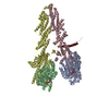
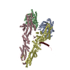
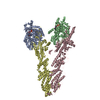
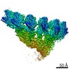
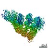
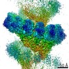
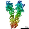
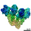
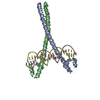
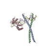
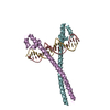
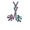
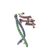
Journal: Elife / Year: 2021 Title: CM1-driven assembly and activation of yeast γ-tubulin small complex underlies microtubule nucleation. Authors: Axel F Brilot / Andrew S Lyon / Alex Zelter / Shruthi Viswanath / Alison Maxwell / Michael J MacCoss / Eric G Muller / Andrej Sali / Trisha N Davis / David A Agard / Abstract: Microtubule (MT) nucleation is regulated by the γ-tubulin ring complex (γTuRC), conserved from yeast to humans. In , γTuRC is composed of seven identical γ-tubulin small complex (γTuSC) sub- ...Microtubule (MT) nucleation is regulated by the γ-tubulin ring complex (γTuRC), conserved from yeast to humans. In , γTuRC is composed of seven identical γ-tubulin small complex (γTuSC) sub-assemblies, which associate helically to template MT growth. γTuRC assembly provides a key point of regulation for the MT cytoskeleton. Here, we combine crosslinking mass spectrometry, X-ray crystallography, and cryo-EM structures of both monomeric and dimeric γTuSCs, and open and closed helical γTuRC assemblies in complex with Spc110p to elucidate the mechanisms of γTuRC assembly. γTuRC assembly is substantially aided by the evolutionarily conserved CM1 motif in Spc110p spanning a pair of adjacent γTuSCs. By providing the highest resolution and most complete views of any γTuSC assembly, our structures allow phosphorylation sites to be mapped, surprisingly suggesting that they are mostly inhibitory. A comparison of our structures with the CM1 binding site in the human γTuRC structure at the interface between GCP2 and GCP6 allows for the interpretation of significant structural changes arising from CM1 helix binding to metazoan γTuRC.
 Movie
Movie Controller


 Open data
 Basic information
 Components
 Keywords
 Function and homology information
 Homo sapiens (human)
 X-RAY DIFFRACTION /
  Authors
 United States, 13items
  Citation
 Structure visualization
 Downloads & links
 Other downloads
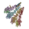
 PDBj
PDBj


 Assembly

 Mass: 18.015 Da / Num. of mol.: 254 / Source method: isolated from a natural source / Formula: H2O
 Sample preparation
 Processing