Switching of origins to a post-replicative state / Activation of the pre-replicative complex / DNA replication preinitiation complex assembly / response to sorbitol / positive regulation of DNA-templated DNA replication / Assembly of the pre-replicative complex / regulation of nuclear cell cycle DNA replication / Orc1 removal from chromatin / negative regulation of DNA-templated DNA replication / DNA replication checkpoint signaling ...Switching of origins to a post-replicative state / Activation of the pre-replicative complex / DNA replication preinitiation complex assembly / response to sorbitol / positive regulation of DNA-templated DNA replication / Assembly of the pre-replicative complex / regulation of nuclear cell cycle DNA replication / Orc1 removal from chromatin / negative regulation of DNA-templated DNA replication / DNA replication checkpoint signaling / attachment of mitotic spindle microtubules to kinetochore / regulation of DNA-templated DNA replication initiation / negative regulation of DNA replication / negative regulation of cell cycle / regulation of DNA replication / transcription repressor complex / DNA polymerase binding / regulation of mitotic cell cycle / positive regulation of DNA replication / animal organ morphogenesis / kinetochore / histone deacetylase binding / transcription corepressor activity / mitotic cell cycle / protein-containing complex assembly / DNA-binding transcription factor binding / nuclear body / negative regulation of DNA-templated transcription / chromatin binding / DNA binding / nucleoplasm / nucleus / cytoplasm / cytosol Similarity search - Function
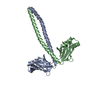
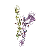
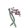
Geminin coiled-coil domain / Geminin/Multicilin / Geminin / CDT1 Geminin-binding domain-like / DNA replication factor Cdt1 / DNA replication factor CDT1 like / DNA replication factor CDT1 like / DNA replication factor Cdt1, C-terminal / DNA replication factor Cdt1, C-terminal WH domain superfamily / DNA replication factor Cdt1 C-terminal domain ...Geminin coiled-coil domain / Geminin/Multicilin / Geminin / CDT1 Geminin-binding domain-like / DNA replication factor Cdt1 / DNA replication factor CDT1 like / DNA replication factor CDT1 like / DNA replication factor Cdt1, C-terminal / DNA replication factor Cdt1, C-terminal WH domain superfamily / DNA replication factor Cdt1 C-terminal domain / Single alpha-helices involved in coiled-coils or other helix-helix interfaces / Winged helix DNA-binding domain superfamily / Up-down Bundle / Mainly Alpha Similarity search - Domain/homology
Method to determine structure: SAD / Resolution: 2.8→30 Å / Cor.coef. Fo:Fc: 0.922 / Cor.coef. Fo:Fc free: 0.898 / SU B: 15.674 / SU ML: 0.297 / Cross valid method: THROUGHOUT / ESU R: 0.738 / ESU R Free: 0.365 / Stereochemistry target values: MAXIMUM LIKELIHOOD / Details: HYDROGENS HAVE BEEN ADDED IN THE RIDING POSITIONS
Rfactor
Num. reflection
% reflection
Selection details
Rfree
0.27737
1432
5.1 %
RANDOM
Rwork
0.23495
-
-
-
obs
0.23715
26561
95.29 %
-
Solvent computation
Ion probe radii: 0.8 Å / Shrinkage radii: 0.8 Å / VDW probe radii: 1.2 Å / Solvent model: MASK
Displacement parameters
Biso mean: 57.505 Å2
Baniso -1
Baniso -2
Baniso -3
1-
-1.8 Å2
0 Å2
1.06 Å2
2-
-
4.54 Å2
0 Å2
3-
-
-
-2.24 Å2
Refinement step
Cycle: LAST / Resolution: 2.8→30 Å
Protein
Nucleic acid
Ligand
Solvent
Total
Num. atoms
5467
0
0
9
5476
Refine LS restraints
Refine-ID
Type
Dev ideal
Dev ideal target
Number
X-RAY DIFFRACTION
r_bond_refined_d
0.014
0.022
5566
X-RAY DIFFRACTION
r_bond_other_d
X-RAY DIFFRACTION
r_angle_refined_deg
1.528
1.968
7495
X-RAY DIFFRACTION
r_angle_other_deg
X-RAY DIFFRACTION
r_dihedral_angle_1_deg
6.378
5
652
X-RAY DIFFRACTION
r_dihedral_angle_2_deg
38.826
24.43
298
X-RAY DIFFRACTION
r_dihedral_angle_3_deg
22.916
15
1078
X-RAY DIFFRACTION
r_dihedral_angle_4_deg
22.492
15
50
X-RAY DIFFRACTION
r_chiral_restr
0.105
0.2
809
X-RAY DIFFRACTION
r_gen_planes_refined
0.005
0.02
4214
X-RAY DIFFRACTION
r_gen_planes_other
X-RAY DIFFRACTION
r_nbd_refined
0.283
0.2
2849
X-RAY DIFFRACTION
r_nbd_other
X-RAY DIFFRACTION
r_nbtor_refined
0.33
0.2
3824
X-RAY DIFFRACTION
r_nbtor_other
X-RAY DIFFRACTION
r_xyhbond_nbd_refined
0.171
0.2
239
X-RAY DIFFRACTION
r_xyhbond_nbd_other
X-RAY DIFFRACTION
r_metal_ion_refined
X-RAY DIFFRACTION
r_metal_ion_other
X-RAY DIFFRACTION
r_symmetry_vdw_refined
0.255
0.2
99
X-RAY DIFFRACTION
r_symmetry_vdw_other
X-RAY DIFFRACTION
r_symmetry_hbond_refined
0.194
0.2
8
X-RAY DIFFRACTION
r_symmetry_hbond_other
X-RAY DIFFRACTION
r_symmetry_metal_ion_refined
X-RAY DIFFRACTION
r_symmetry_metal_ion_other
X-RAY DIFFRACTION
r_mcbond_it
0.944
1.5
3400
X-RAY DIFFRACTION
r_mcbond_other
X-RAY DIFFRACTION
r_mcangle_it
1.448
2
5378
X-RAY DIFFRACTION
r_scbond_it
2.064
3
2401
X-RAY DIFFRACTION
r_scangle_it
3.522
4.5
2117
X-RAY DIFFRACTION
r_rigid_bond_restr
X-RAY DIFFRACTION
r_sphericity_free
X-RAY DIFFRACTION
r_sphericity_bonded
LS refinement shell
Resolution: 2.8→2.872 Å / Total num. of bins used: 20
Rfactor
Num. reflection
% reflection
Rfree
0.464
72
-
Rwork
0.392
1356
-
obs
-
-
66.92 %
+
About Yorodumi
-
News
-
Feb 9, 2022. New format data for meta-information of EMDB entries
New format data for meta-information of EMDB entries
Version 3 of the EMDB header file is now the official format.
The previous official version 1.9 will be removed from the archive.
In the structure databanks used in Yorodumi, some data are registered as the other names, "COVID-19 virus" and "2019-nCoV". Here are the details of the virus and the list of structure data.
Jan 31, 2019. EMDB accession codes are about to change! (news from PDBe EMDB page)
EMDB accession codes are about to change! (news from PDBe EMDB page)
The allocation of 4 digits for EMDB accession codes will soon come to an end. Whilst these codes will remain in use, new EMDB accession codes will include an additional digit and will expand incrementally as the available range of codes is exhausted. The current 4-digit format prefixed with “EMD-” (i.e. EMD-XXXX) will advance to a 5-digit format (i.e. EMD-XXXXX), and so on. It is currently estimated that the 4-digit codes will be depleted around Spring 2019, at which point the 5-digit format will come into force.
The EM Navigator/Yorodumi systems omit the EMD- prefix.
Related info.:Q: What is EMD? / ID/Accession-code notation in Yorodumi/EM Navigator
Yorodumi is a browser for structure data from EMDB, PDB, SASBDB, etc.
This page is also the successor to EM Navigator detail page, and also detail information page/front-end page for Omokage search.
The word "yorodu" (or yorozu) is an old Japanese word meaning "ten thousand". "mi" (miru) is to see.
Related info.:EMDB / PDB / SASBDB / Comparison of 3 databanks / Yorodumi Search / Aug 31, 2016. New EM Navigator & Yorodumi / Yorodumi Papers / Jmol/JSmol / Function and homology information / Changes in new EM Navigator and Yorodumi
 Movie
Movie Controller


 Open data
 Basic information
 Components
 Keywords
 Function and homology information
 X-RAY DIFFRACTION /
  Authors
 Citation
 Structure visualization
 Downloads & links
 Other downloads
 PDBj
PDBj
 Assembly


 Mass: 18.015 Da / Num. of mol.: 9 / Source method: isolated from a natural source / Formula: H2O
 Sample preparation
 / Beamline: 19-BM / Wavelength: 0.9792 Å
 Processing