- EMDB-23639: Single-particle reconstruction of a dimer of the Yeast gamma-TuSC -
+
Open data
ID or keywords:
Loading...
-
Basic information
Entry
Database: EMDB / ID: EMD-23639
Title
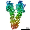
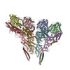
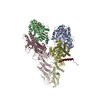
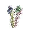
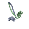
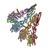
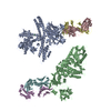
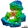
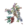
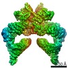
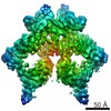
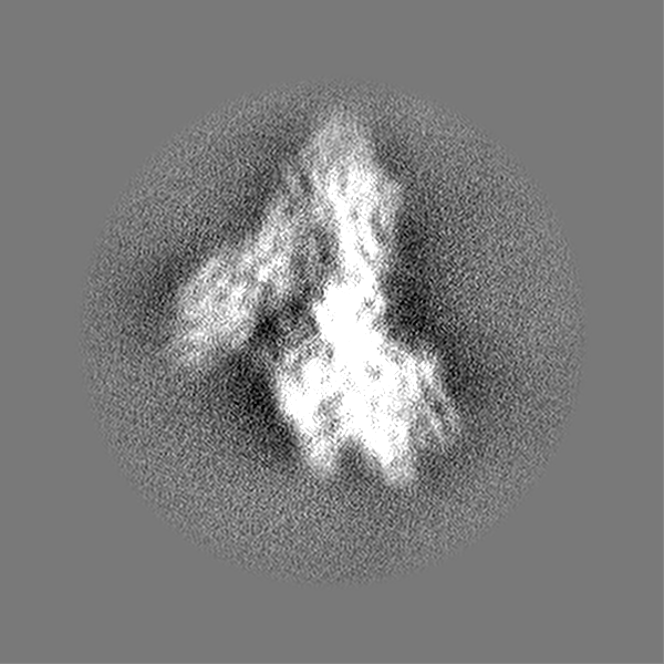
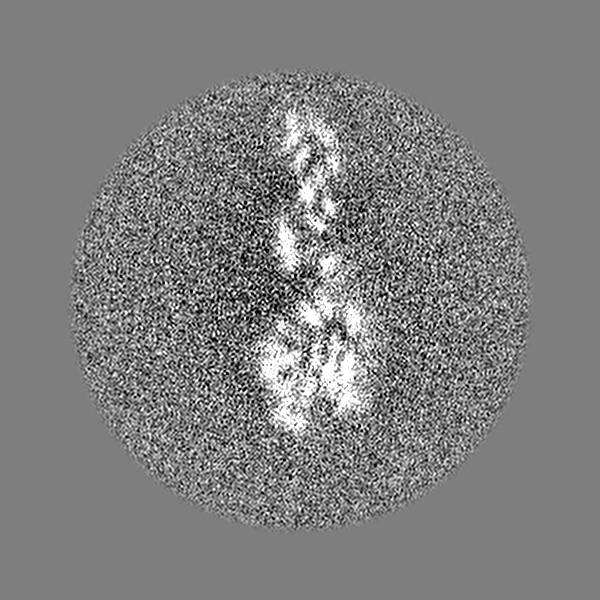
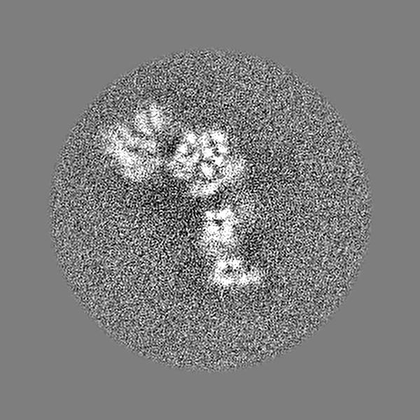
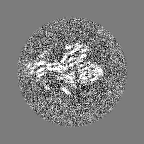
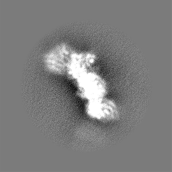
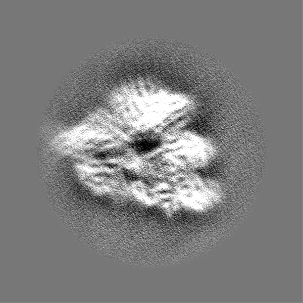
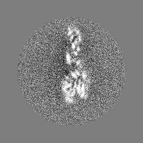
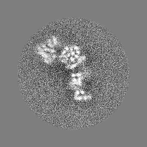
Single-particle reconstruction of a dimer of the Yeast gamma-TuSC
Map data
Single-particle reconstruction of a dimer of the Yeast gamma-TuSC half map 1
Sample
Complex: Single-particle reconstruction of a dimer of the Yeast gamma-TuSC
Protein or peptide: Tub4p (gamma-tubulin)
Protein or peptide: Spc97p
Protein or peptide: Spc98p
Function / homology
Function and homology information
gamma-tubulin complex localization to nuclear side of mitotic spindle pole body / protein localization to mitotic spindle pole body / inner plaque of spindle pole body / microtubule nucleation by spindle pole body / outer plaque of spindle pole body / gamma-tubulin small complex / central plaque of spindle pole body / karyogamy involved in conjugation with cellular fusion / regulation of microtubule nucleation / microtubule nucleator activity ...gamma-tubulin complex localization to nuclear side of mitotic spindle pole body / protein localization to mitotic spindle pole body / inner plaque of spindle pole body / microtubule nucleation by spindle pole body / outer plaque of spindle pole body / gamma-tubulin small complex / central plaque of spindle pole body / karyogamy involved in conjugation with cellular fusion / regulation of microtubule nucleation / microtubule nucleator activity / mitotic spindle elongation / mitotic spindle pole body / gamma-tubulin complex / gamma-tubulin ring complex / meiotic spindle organization / positive regulation of microtubule nucleation / microtubule nucleation / spindle pole body / gamma-tubulin binding / positive regulation of cytoplasmic translation / mitotic sister chromatid segregation / spindle assembly / cytoplasmic microtubule organization / mitotic spindle organization / meiotic cell cycle / structural constituent of cytoskeleton / spindle / spindle pole / mitotic cell cycle / protein-containing complex assembly / cytoskeleton / microtubule / calmodulin binding / GTP binding / protein-containing complex binding / nucleus / cytoplasm Similarity search - Function
Spindle pole body component 110 / Spindle pole body component SPC97 / Tubulin gamma chain / Spindle pole body component SPC98 Similarity search - Component
Biological species
Saccharomyces cerevisiae (brewer's yeast)
Method
single particle reconstruction / cryo EM / Resolution: 4.45 Å
National Institutes of Health/National Institute of General Medical Sciences (NIH/NIGMS)
R01 GM031627
United States
National Institutes of Health/National Institute of General Medical Sciences (NIH/NIGMS)
R35 GM118099
United States
National Institutes of Health/National Institute of General Medical Sciences (NIH/NIGMS)
P01 GM105537
United States
National Institutes of Health/National Institute of General Medical Sciences (NIH/NIGMS)
P41 GM103533
United States
National Institutes of Health/National Institute of General Medical Sciences (NIH/NIGMS)
GM083960
United States
National Institutes of Health/National Institute of General Medical Sciences (NIH/NIGMS)
GM109824
United States
National Science Foundation (NSF, United States)
1144247
United States
National Institutes of Health/Office of the Director
1S10OD020054
United States
National Institutes of Health/Office of the Director
1S10OD021741
United States
National Institutes of Health/National Institute of General Medical Sciences (NIH/NIGMS)
R01 GM124149
United States
National Institutes of Health/National Institute of General Medical Sciences (NIH/NIGMS)
P30 GM124169
United States
Department of Energy (DOE, United States)
DE-AC02-05CH11231
United States
Citation
Journal: Elife / Year: 2021 Title: CM1-driven assembly and activation of yeast γ-tubulin small complex underlies microtubule nucleation. Authors: Axel F Brilot / Andrew S Lyon / Alex Zelter / Shruthi Viswanath / Alison Maxwell / Michael J MacCoss / Eric G Muller / Andrej Sali / Trisha N Davis / David A Agard / Abstract: Microtubule (MT) nucleation is regulated by the γ-tubulin ring complex (γTuRC), conserved from yeast to humans. In , γTuRC is composed of seven identical γ-tubulin small complex (γTuSC) sub- ...Microtubule (MT) nucleation is regulated by the γ-tubulin ring complex (γTuRC), conserved from yeast to humans. In , γTuRC is composed of seven identical γ-tubulin small complex (γTuSC) sub-assemblies, which associate helically to template MT growth. γTuRC assembly provides a key point of regulation for the MT cytoskeleton. Here, we combine crosslinking mass spectrometry, X-ray crystallography, and cryo-EM structures of both monomeric and dimeric γTuSCs, and open and closed helical γTuRC assemblies in complex with Spc110p to elucidate the mechanisms of γTuRC assembly. γTuRC assembly is substantially aided by the evolutionarily conserved CM1 motif in Spc110p spanning a pair of adjacent γTuSCs. By providing the highest resolution and most complete views of any γTuSC assembly, our structures allow phosphorylation sites to be mapped, surprisingly suggesting that they are mostly inhibitory. A comparison of our structures with the CM1 binding site in the human γTuRC structure at the interface between GCP2 and GCP6 allows for the interpretation of significant structural changes arising from CM1 helix binding to metazoan γTuRC.
History
Deposition
Mar 17, 2021
-
Header (metadata) release
May 12, 2021
-
Map release
May 12, 2021
-
Update
May 19, 2021
-
Current status
May 19, 2021
Processing site: RCSB / Status: Released
-
Structure visualization
Movie
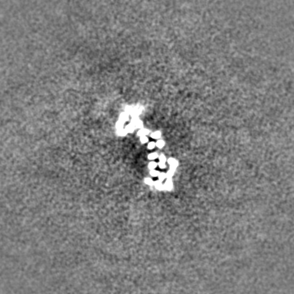
Surface view with section colored by density value
Specimen holder model: OTHER / Cooling holder cryogen: NITROGEN
Experimental equipment
Model: Tecnai Polara / Image courtesy: FEI Company
+
Image processing
Particle selection
Number selected: 3210917 / Details: cisTEM picking
CTF correction
Software - Name: cisTEM (ver. 1.0 beta) / Details: Final reconstruction in cisTEM
Startup model
Type of model: INSILICO MODEL In silico model: Particle coordinates were semi-automatically picked from filtered and binned images using the e2boxer swarm tool (Tang et al., 2007). Particles were extracted using Relion (Scheres, ...In silico model: Particle coordinates were semi-automatically picked from filtered and binned images using the e2boxer swarm tool (Tang et al., 2007). Particles were extracted using Relion (Scheres, 2012) with a box size of 384 physical pixels resampled to 96 pixels for initial processing. A dataset of ~50,000 particles from 217 micrographs was used to generate 300 2D classes using Relion 1.3. 23 classes were selected and used in the generation of a gamma-TuSC monomer initial model using the e2initialmodel.py function in EMAN2.
Final reconstruction
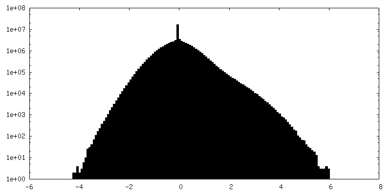
Number classes used: 1 / Applied symmetry - Point group: C1 (asymmetric) / Algorithm: FOURIER SPACE / Resolution.type: BY AUTHOR / Resolution: 4.45 Å / Resolution method: FSC 0.143 CUT-OFF / Software - Name: cisTEM (ver. 1.0 Beta) / Number images used: 506014
In the structure databanks used in Yorodumi, some data are registered as the other names, "COVID-19 virus" and "2019-nCoV". Here are the details of the virus and the list of structure data.
Jan 31, 2019. EMDB accession codes are about to change! (news from PDBe EMDB page)
EMDB accession codes are about to change! (news from PDBe EMDB page)
The allocation of 4 digits for EMDB accession codes will soon come to an end. Whilst these codes will remain in use, new EMDB accession codes will include an additional digit and will expand incrementally as the available range of codes is exhausted. The current 4-digit format prefixed with “EMD-” (i.e. EMD-XXXX) will advance to a 5-digit format (i.e. EMD-XXXXX), and so on. It is currently estimated that the 4-digit codes will be depleted around Spring 2019, at which point the 5-digit format will come into force.
The EM Navigator/Yorodumi systems omit the EMD- prefix.
Related info.:Q: What is EMD? / ID/Accession-code notation in Yorodumi/EM Navigator
Yorodumi is a browser for structure data from EMDB, PDB, SASBDB, etc.
This page is also the successor to EM Navigator detail page, and also detail information page/front-end page for Omokage search.
The word "yorodu" (or yorozu) is an old Japanese word meaning "ten thousand". "mi" (miru) is to see.
Related info.:EMDB / PDB / SASBDB / Comparison of 3 databanks / Yorodumi Search / Aug 31, 2016. New EM Navigator & Yorodumi / Yorodumi Papers / Jmol/JSmol / Function and homology information / Changes in new EM Navigator and Yorodumi
 Movie
Movie Controller

 Yorodumi
 Open data
 Basic information
 Map data
 Sample
 Function and homology information
 Authors
 United States, 13 items
  Citation
 Structure visualization
 Downloads & links
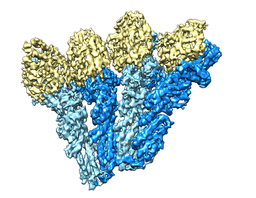 emd_23639.png
 http://ftp.pdbj.org/pub/emdb/structures/EMD-23639


 Z (Sec.)
 Y (Row.)
 X (Col.)


 Sample components
 Spodoptera frugiperda (fall armyworm) / Recombinant strain: SF9
 Processing
 Electron microscopy
 FIELD EMISSION GUN
