Movie
Movie Controller


+ Open data

- Basic information
| Entry | Database: EMDB / ID: EMD-6399 | |||||||||
|---|---|---|---|---|---|---|---|---|---|---|
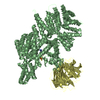
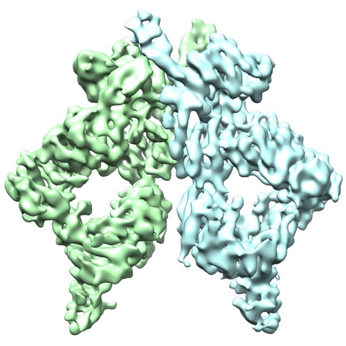
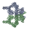
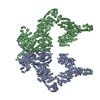
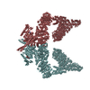
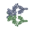
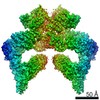
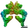
| Title | Structure of the intact ATM/Tel1 kinase | |||||||||
 Map data Map data | Reconstruction of ATM/Tel1 | |||||||||
 Sample Sample |
| |||||||||
 Keywords Keywords | ATM/Tel1 / DSB / kinase activity | |||||||||
| Biological species |  | |||||||||
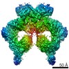
| Method | single particle reconstruction / cryo EM / negative staining / Resolution: 8.7 Å | |||||||||
 Authors Authors | Wang XJ / Chu HY / Lv MJ / Zhang ZH / Qiu SW / Liu HY / Shen XT / Wang WW / Cai G | |||||||||
 Citation Citation | Journal: Nat Commun / Year: 2016 Title: Structure of the intact ATM/Tel1 kinase. Authors: Xuejuan Wang / Huanyu Chu / Mengjuan Lv / Zhihui Zhang / Shuwan Qiu / Haiyan Liu / Xuetong Shen / Weiwu Wang / Gang Cai /   Abstract: The ataxia-telangiectasia mutated (ATM) protein is an apical kinase that orchestrates the multifaceted DNA-damage response. Normally, ATM kinase is in an inactive, homodimer form and is transformed ...The ataxia-telangiectasia mutated (ATM) protein is an apical kinase that orchestrates the multifaceted DNA-damage response. Normally, ATM kinase is in an inactive, homodimer form and is transformed into monomers upon activation. Besides a conserved kinase domain at the C terminus, ATM contains three other structural modules, referred to as FAT, FATC and N-terminal helical solenoid. Here we report the first cryo-EM structure of ATM kinase, which is an intact homodimeric ATM/Tel1 from Schizosaccharomyces pombe. We show that two monomers directly contact head-to-head through the FAT and kinase domains. The tandem N-terminal helical solenoid tightly packs against the FAT and kinase domains. The structure suggests that ATM/Tel1 dimer interface and the consecutive HEAT repeats inhibit the binding of kinase substrates and regulators by steric hindrance. Our study provides a structural framework for understanding the mechanisms of ATM/Tel1 regulation as well as the development of new therapeutic agents. | |||||||||
| History |
|
- Structure visualization
| Movie |
 Movie viewer Movie viewer |
|---|---|
| Structure viewer | EM map: SurfViewMolmilJmol/JSmol |
| Supplemental images |
- Downloads & links
-EMDB archive
| Map data | emd_6399.map.gz | 59.6 MB | EMDB map data format | |
|---|---|---|---|---|
| Header (meta data) | emd-6399-v30.xmlemd-6399.xml | 10.7 KB 10.7 KB | Display Display | EMDB header |
| Images | emd_6399.tif | 762.2 KB | ||
| Archive directory |  http://ftp.pdbj.org/pub/emdb/structures/EMD-6399ftp://ftp.pdbj.org/pub/emdb/structures/EMD-6399 http://ftp.pdbj.org/pub/emdb/structures/EMD-6399ftp://ftp.pdbj.org/pub/emdb/structures/EMD-6399 | HTTPS FTP |
-Related structure data
| Similar structure data |
|---|

-Links
| EMDB pages | EMDB (EBI/PDBe) / EMDataResource |
|---|
-Map
| File | Download / File: emd_6399.map.gz / Format: CCP4 / Size: 62.5 MB / Type: IMAGE STORED AS FLOATING POINT NUMBER (4 BYTES) | ||||||||||||||||||||||||||||||||||||||||||||||||||||||||||||
|---|---|---|---|---|---|---|---|---|---|---|---|---|---|---|---|---|---|---|---|---|---|---|---|---|---|---|---|---|---|---|---|---|---|---|---|---|---|---|---|---|---|---|---|---|---|---|---|---|---|---|---|---|---|---|---|---|---|---|---|---|---|
| Annotation | Reconstruction of ATM/Tel1 | ||||||||||||||||||||||||||||||||||||||||||||||||||||||||||||
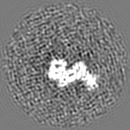
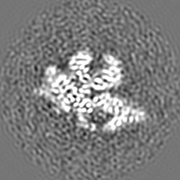
| Projections & slices | Image control
Images are generated by Spider. | ||||||||||||||||||||||||||||||||||||||||||||||||||||||||||||
| Voxel size | X=Y=Z: 1.42 Å | ||||||||||||||||||||||||||||||||||||||||||||||||||||||||||||
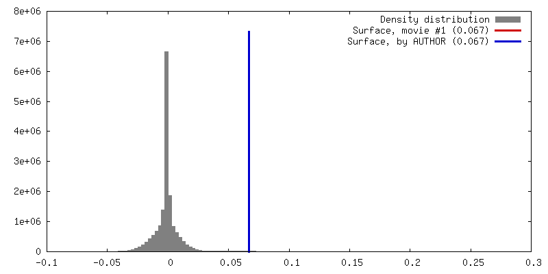
| Density |
| ||||||||||||||||||||||||||||||||||||||||||||||||||||||||||||
| Symmetry | Space group: 1 | ||||||||||||||||||||||||||||||||||||||||||||||||||||||||||||
| Details | EMDB XML:
CCP4 map header:
| ||||||||||||||||||||||||||||||||||||||||||||||||||||||||||||
 Z (Sec.)
 Y (Row.)
 X (Col.)


-Supplemental data
- Sample components
-Entire : Endogenous ATM/Tel1 purified directly from the yeast cells
| Entire | Name: Endogenous ATM/Tel1 purified directly from the yeast cells |
|---|---|
| Components |
|
-Supramolecule #1000: Endogenous ATM/Tel1 purified directly from the yeast cells
| Supramolecule | Name: Endogenous ATM/Tel1 purified directly from the yeast cells type: sample / ID: 1000 Oligomeric state: An asymmetric homo-dimeric architecture with pseudo C2 symmetry Number unique components: 1 |
|---|---|
| Molecular weight | Experimental: 700 KDa / Theoretical: 700 KDa / Method: Sedimentation |
-Macromolecule #1: ATM/Tel1
| Macromolecule | Name: ATM/Tel1 / type: protein_or_peptide / ID: 1 / Number of copies: 2 / Oligomeric state: Dimer / Recombinant expression: No / Database: NCBI |
|---|---|
| Source (natural) | Organism: |
| Molecular weight | Experimental: 700 KDa / Theoretical: 700 KDa |
-Experimental details
-Structure determination
| Method | negative staining, cryo EM |
|---|---|
 Processing Processing | single particle reconstruction |
| Aggregation state | particle |
-Sample preparation
| Concentration | 0.1 mg/mL |
|---|---|
| Buffer | pH: 7.6 Details: 50mmol/L Tris, 100mmol/L ammonium sulfate, 10%(v/v) glycerol, 1mmol/L EDTA, 10umol/L ZnSO4, 0.02% NP-40, 10mmol/b-ME |
| Staining | Type: NEGATIVE Details: Sample was applied to a carbon-coated 400-mesh Cu EM specimen grid freshly glow discharged and was then preserved by staining with 0.75%(w/w) uranyl formate solution. |
| Grid | Details: a carbon-coated 400-mesh Cu EM specimen grid freshly glow discharged |
| Vitrification | Cryogen name: ETHANE / Chamber humidity: 100 % / Chamber temperature: 120 K / Instrument: FEI VITROBOT MARK IV Timed resolved state: Vitrified 30 msec after spraying with effector Method: Blot for 3-4 seconds before plunging |
- Electron microscopy
| Microscope | FEI TITAN KRIOS |
|---|---|
| Alignment procedure | Legacy - Astigmatism: Objective lens astigmatism was corrected at 150,000 times magnification |
| Date | Mar 3, 2015 |
| Image recording | Category: CCD / Film or detector model: FEI FALCON II (4k x 4k) / Average electron dose: 35 e/Å2 |
| Electron beam | Acceleration voltage: 300 kV / Electron source:  FIELD EMISSION GUN FIELD EMISSION GUN |
| Electron optics | Illumination mode: FLOOD BEAM / Imaging mode: BRIGHT FIELD / Cs: 2.7 mm / Nominal defocus max: 4.0 µm / Nominal defocus min: 2.5 µm / Nominal magnification: 59000 |
| Sample stage | Specimen holder model: FEI TITAN KRIOS AUTOGRID HOLDER |
| Experimental equipment |  Model: Titan Krios / Image courtesy: FEI Company |
-Image processing
| Details | The particles were selected using a semi-automated procedure in RELION. |
|---|---|
| CTF correction | Details: Each micrograph |
| Final reconstruction | Algorithm: OTHER / Resolution.type: BY AUTHOR / Resolution: 8.7 Å / Resolution method: OTHER / Software - Name: RELION / Number images used: 57435 |
| Final two d classification | Number classes: 6 |
-Atomic model buiding 1
| Initial model | PDB ID: |
|---|---|
| Software | Name: Chimera |
| Refinement | Space: REAL / Protocol: RIGID BODY FIT |