positive regulation of ERK5 cascade / ASP-3026-resistant ALK mutants / NVP-TAE684-resistant ALK mutants / alectinib-resistant ALK mutants / brigatinib-resistant ALK mutants / ceritinib-resistant ALK mutants / crizotinib-resistant ALK mutants / lorlatinib-resistant ALK mutants / MDK and PTN in ALK signaling / receptor signaling protein tyrosine kinase activator activity ...positive regulation of ERK5 cascade / ASP-3026-resistant ALK mutants / NVP-TAE684-resistant ALK mutants / alectinib-resistant ALK mutants / brigatinib-resistant ALK mutants / ceritinib-resistant ALK mutants / crizotinib-resistant ALK mutants / lorlatinib-resistant ALK mutants / MDK and PTN in ALK signaling / receptor signaling protein tyrosine kinase activator activity / regulation of dopamine receptor signaling pathway / response to environmental enrichment / ALK mutants bind TKIs / peptidyl-tyrosine autophosphorylation / phosphorylation / Signaling by LTK / positive regulation of dendrite development / regulation of neuron differentiation / Signaling by ALK / adult behavior / response to stress / negative regulation of lipid catabolic process / neuron development / energy homeostasis / swimming behavior / transmembrane receptor protein tyrosine kinase activity / cell surface receptor protein tyrosine kinase signaling pathway / cytokine activity / : / hippocampus development / receptor protein-tyrosine kinase / positive regulation of neuron projection development / receptor tyrosine kinase binding / Signaling by ALK fusions and activated point mutants / protein autophosphorylation / regulation of cell population proliferation / heparin binding / protein tyrosine kinase activity / regulation of apoptotic process / positive regulation of ERK1 and ERK2 cascade / signaling receptor complex / signal transduction / protein-containing complex / : / extracellular exosome / extracellular region / ATP binding / identical protein binding / plasma membrane Similarity search - Function
ALK and LTK ligand 1/2 / ALK and LTK ligand 1/2 / : / ALK/LTK, Glycine-rich domain / MAM domain, meprin/A5/mu / MAM domain / MAM domain profile. / Low-density lipoprotein receptor domain class A / Tyrosine-protein kinase, receptor class II, conserved site / Receptor tyrosine kinase class II signature. ...ALK and LTK ligand 1/2 / ALK and LTK ligand 1/2 / : / ALK/LTK, Glycine-rich domain / MAM domain, meprin/A5/mu / MAM domain / MAM domain profile. / Low-density lipoprotein receptor domain class A / Tyrosine-protein kinase, receptor class II, conserved site / Receptor tyrosine kinase class II signature. / Low-density lipoprotein (LDL) receptor class A repeat / LDL receptor-like superfamily / : / Tyrosine-protein kinase, catalytic domain / Tyrosine kinase, catalytic domain / Tyrosine protein kinases specific active-site signature. / Tyrosine-protein kinase, active site / Concanavalin A-like lectin/glucanase domain superfamily / Protein tyrosine and serine/threonine kinase / Serine-threonine/tyrosine-protein kinase, catalytic domain / Protein kinase, ATP binding site / Protein kinases ATP-binding region signature. / Protein kinase domain profile. / Protein kinase domain / Protein kinase-like domain superfamily Similarity search - Domain/homology
In the structure databanks used in Yorodumi, some data are registered as the other names, "COVID-19 virus" and "2019-nCoV". Here are the details of the virus and the list of structure data.
Jan 31, 2019. EMDB accession codes are about to change! (news from PDBe EMDB page)
EMDB accession codes are about to change! (news from PDBe EMDB page)
The allocation of 4 digits for EMDB accession codes will soon come to an end. Whilst these codes will remain in use, new EMDB accession codes will include an additional digit and will expand incrementally as the available range of codes is exhausted. The current 4-digit format prefixed with “EMD-” (i.e. EMD-XXXX) will advance to a 5-digit format (i.e. EMD-XXXXX), and so on. It is currently estimated that the 4-digit codes will be depleted around Spring 2019, at which point the 5-digit format will come into force.
The EM Navigator/Yorodumi systems omit the EMD- prefix.
Related info.:Q: What is EMD? / ID/Accession-code notation in Yorodumi/EM Navigator
Yorodumi is a browser for structure data from EMDB, PDB, SASBDB, etc.
This page is also the successor to EM Navigator detail page, and also detail information page/front-end page for Omokage search.
The word "yorodu" (or yorozu) is an old Japanese word meaning "ten thousand". "mi" (miru) is to see.
Related info.:EMDB / PDB / SASBDB / Comparison of 3 databanks / Yorodumi Search / Aug 31, 2016. New EM Navigator & Yorodumi / Yorodumi Papers / Jmol/JSmol / Function and homology information / Changes in new EM Navigator and Yorodumi
 Movie
Movie Controller


 Open data
 Basic information
 Components
 Keywords
 Function and homology information
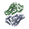
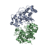
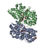
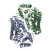
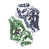
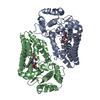
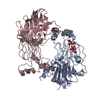
 Homo sapiens (human)
 X-RAY DIFFRACTION /
  Authors
 United States, 2items
  Citation
 Structure visualization
 Downloads & links
 Other downloads


 PDBj
PDBj


 Assembly
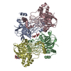
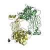
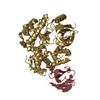
 Trichoplusia ni (cabbage looper)
 Mass: 192.124 Da / Num. of mol.: 2 / Source method: obtained synthetically / Formula: C6H8O7
 Type: D-saccharide, beta linking / Mass: 221.208 Da / Num. of mol.: 1 / Source method: obtained synthetically / Formula: C8H15NO6
 Sample preparation
 Processing