 Movie
Movie Controller

[English] 日本語
 Yorodumi
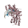
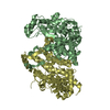
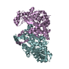
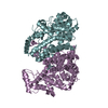
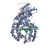
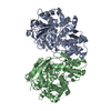
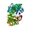
- PDB-1mc8: Crystal Structure of Flap Endonuclease-1 R42E mutant from Pyrococ... -
+ Open data

- Basic information
| Entry | Database: PDB / ID: 1mc8 | ||||||
|---|---|---|---|---|---|---|---|
| Title | Crystal Structure of Flap Endonuclease-1 R42E mutant from Pyrococcus horikoshii | ||||||
 Components Components | Flap Endonuclease-1 | ||||||
 Keywords Keywords | HYDROLASE / flexible loop | ||||||
| Function / homology |  Function and homology information Function and homology information5'-flap endonuclease activity / DNA replication, removal of RNA primer / 5'-3' exonuclease activity / Hydrolases; Acting on ester bonds / DNA repair / magnesium ion binding / DNA binding Similarity search - Function | ||||||
| Biological species |   Pyrococcus horikoshii (archaea) Pyrococcus horikoshii (archaea) | ||||||
| Method |  X-RAY DIFFRACTION / SYNCHROTRON / MOLECULAR REPLACEMENT / Resolution: 3.1 Å X-RAY DIFFRACTION / SYNCHROTRON / MOLECULAR REPLACEMENT / Resolution: 3.1 Å | ||||||
 Authors Authors | Matsui, E. / Musti, K.V. / Abe, J. / Yamazaki, K. / Matsui, I. / Harata, K. | ||||||
 Citation Citation | Journal: J.BIOL.CHEM. / Year: 2002 Title: Molecular Structure and Novel DNA Binding Sites Located in Loops of Flap Endonuclease-1 from Pyrococcus horikoshii Authors: Matsui, E. / Musti, K.V. / Abe, J. / Yamazaki, K. / Matsui, I. / Harata, K. | ||||||
| History |
|
- Structure visualization
| Structure viewer | Molecule: MolmilJmol/JSmol |
|---|
- Downloads & links
-Download
| PDBx/mmCIF format | 1mc8.cif.gz | 139.8 KB | Display | PDBx/mmCIF format |
|---|---|---|---|---|
| PDB format | pdb1mc8.ent.gz | 111.1 KB | Display | PDB format |
| PDBx/mmJSON format | 1mc8.json.gz | Tree view | PDBx/mmJSON format | |
| Others |  Other downloads Other downloads |
-Validation report
| Arichive directory | https://data.pdbj.org/pub/pdb/validation_reports/mc/1mc8ftp://data.pdbj.org/pub/pdb/validation_reports/mc/1mc8 | HTTPS FTP |
|---|
-Related structure data
| Related structure data |  1a76S S: Starting model for refinement |
|---|---|
| Similar structure data |


-Links
 PDBj
PDBj

- Assembly
| Deposited unit | 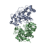
| ||||||||
|---|---|---|---|---|---|---|---|---|---|
| 1 |
| ||||||||
| Unit cell |
|
-Components
| #1: Protein | Mass: 38980.082 Da / Num. of mol.: 2 / Mutation: R42E Source method: isolated from a genetically manipulated source Source: (gene. exp.) Pyrococcus horikoshii (archaea) / Plasmid: pET-11a / Production host:  |
|---|
-Experimental details
-Experiment
| Experiment | Method: X-RAY DIFFRACTION / Number of used crystals: 1 |
|---|
- Sample preparation
| Crystal | Density Matthews: 2.63 Å3/Da / Density % sol: 53.17 % | ||||||||||||||||||||||||||||||||||||||||||
|---|---|---|---|---|---|---|---|---|---|---|---|---|---|---|---|---|---|---|---|---|---|---|---|---|---|---|---|---|---|---|---|---|---|---|---|---|---|---|---|---|---|---|---|
| Crystal grow | Temperature: 298 K / Method: vapor diffusion, hanging drop / pH: 6 Details: PEG6000, sodium chloride, pH 6.0, VAPOR DIFFUSION, HANGING DROP, temperature 298K | ||||||||||||||||||||||||||||||||||||||||||
| Crystal grow | *PLUS | ||||||||||||||||||||||||||||||||||||||||||
| Components of the solutions | *PLUS
|
-Data collection
| Diffraction | Mean temperature: 298 K |
|---|---|
| Diffraction source | Source: SYNCHROTRON / Site: Photon Factory  / Beamline: BL-6A / Wavelength: 1 Å / Beamline: BL-6A / Wavelength: 1 Å |
| Detector | Type: ADSC QUANTUM 4 / Detector: CCD / Date: Nov 16, 2000 |
| Radiation | Monochromator: Si(111) / Protocol: SINGLE WAVELENGTH / Monochromatic (M) / Laue (L): M / Scattering type: x-ray |
| Radiation wavelength | Wavelength: 1 Å / Relative weight: 1 |
| Reflection | Resolution: 3→34.7 Å / Num. all: 15903 / Num. obs: 15903 / % possible obs: 100 % / Observed criterion σ(F): 0 / Observed criterion σ(I): 0 / Redundancy: 5.7 % / Rmerge(I) obs: 0.104 |
| Reflection shell | Resolution: 3→3.16 Å / Redundancy: 5.6 % / Rmerge(I) obs: 0.494 / % possible all: 98.6 |
| Reflection | *PLUS % possible obs: 100 % / Num. measured all: 135478 / Rmerge(I) obs: 0.104 |
| Reflection shell | *PLUS Rmerge(I) obs: 0.494 |
- Processing
| Software |
| |||||||||||||||||||||
|---|---|---|---|---|---|---|---|---|---|---|---|---|---|---|---|---|---|---|---|---|---|---|
| Refinement | Method to determine structure: MOLECULAR REPLACEMENT Starting model: PDB entry 1A76 Resolution: 3.1→8 Å / Isotropic thermal model: Isotropic / σ(F): 2 / Stereochemistry target values: Engh & Huber
| |||||||||||||||||||||
| Displacement parameters | Biso mean: 37.8 Å2 | |||||||||||||||||||||
| Refinement step | Cycle: LAST / Resolution: 3.1→8 Å
| |||||||||||||||||||||
| Refine LS restraints |
| |||||||||||||||||||||
| LS refinement shell | Resolution: 3.1→3.15 Å
| |||||||||||||||||||||
| Refinement | *PLUS Highest resolution: 3.1 Å / Lowest resolution: 8 Å / Rfactor obs: 0.197 / Rfactor Rfree: 0.279 / Rfactor Rwork: 0.19 | |||||||||||||||||||||
| Solvent computation | *PLUS | |||||||||||||||||||||
| Displacement parameters | *PLUS | |||||||||||||||||||||
| Refine LS restraints | *PLUS
| |||||||||||||||||||||
| LS refinement shell | *PLUS Rfactor Rfree: 0.458 / Rfactor Rwork: 0.328 |
