 Movie
Movie Controller

[English] 日本語
 Yorodumi
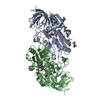
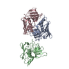
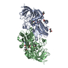
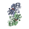
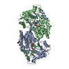
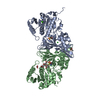
- PDB-4kp2: Crystal structure of homoaconitase large subunit from methanococc... -
+ Open data

- Basic information
| Entry | Database: PDB / ID: 4kp2 | ||||||
|---|---|---|---|---|---|---|---|
| Title | Crystal structure of homoaconitase large subunit from methanococcus jannaschii (MJ1003) | ||||||
 Components Components | Homoaconitase large subunit | ||||||
 Keywords Keywords | LYASE / Aconitase family / alpha-beta-alpha 3-layer sandwich / Isomerase / iron-sulfur cluster binding / small subunit (MJ1271) binding | ||||||
| Function / homology |  Function and homology information Function and homology informationmethanogen homoaconitase / homoaconitate hydratase activity / maleate hydratase activity / 3-isopropylmalate dehydratase activity / L-leucine biosynthetic process / 4 iron, 4 sulfur cluster binding / metal ion binding Similarity search - Function | ||||||
| Biological species |   Methanocaldococcus jannaschii (archaea) Methanocaldococcus jannaschii (archaea) | ||||||
| Method |  X-RAY DIFFRACTION / SYNCHROTRON / MOLECULAR REPLACEMENT / Resolution: 2.504 Å X-RAY DIFFRACTION / SYNCHROTRON / MOLECULAR REPLACEMENT / Resolution: 2.504 Å | ||||||
 Authors Authors | Hwang, K.Y. / Lee, E.H. | ||||||
 Citation Citation | Journal: Acta Crystallogr.,Sect.D / Year: 2014 Title: Structural characterization and comparison of the large subunits of IPM isomerase and homoaconitase from Methanococcus jannaschii Authors: Lee, E.H. / Lee, K. / Hwang, K.Y. | ||||||
| History |
|
- Structure visualization
| Structure viewer | Molecule: MolmilJmol/JSmol |
|---|
- Downloads & links
-Download
| PDBx/mmCIF format | 4kp2.cif.gz | 168.9 KB | Display | PDBx/mmCIF format |
|---|---|---|---|---|
| PDB format | pdb4kp2.ent.gz | 132.3 KB | Display | PDB format |
| PDBx/mmJSON format | 4kp2.json.gz | Tree view | PDBx/mmJSON format | |
| Others |  Other downloads Other downloads |
-Validation report
| Arichive directory | https://data.pdbj.org/pub/pdb/validation_reports/kp/4kp2ftp://data.pdbj.org/pub/pdb/validation_reports/kp/4kp2 | HTTPS FTP |
|---|
-Related structure data
| Related structure data |  4kp1SC 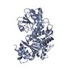 4nqyC S: Starting model for refinement C: citing same article ( |
|---|---|
| Similar structure data |


-Links
 PDBj
PDBj

- Assembly
| Deposited unit | 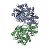
| ||||||||
|---|---|---|---|---|---|---|---|---|---|
| 1 | 
| ||||||||
| Unit cell |
|
-Components
| #1: Protein | Mass: 48427.609 Da / Num. of mol.: 2 Source method: isolated from a genetically manipulated source Source: (gene. exp.) Methanocaldococcus jannaschii (archaea)Strain: ATCC 43067 / DSM 2661 / JAL-1 / JCM 10045 / NBRC 100440 Gene: hacA, MJ1003 / Plasmid: pET28a / Production host:  References: UniProt: Q58409, EC: 4.2.1.36, Lyases; Carbon-oxygen lyases; Hydro-lyases #2: Water | ChemComp-HOH / |  Mass: 18.015 Da / Num. of mol.: 52 / Source method: isolated from a natural source / Formula: H2O Mass: 18.015 Da / Num. of mol.: 52 / Source method: isolated from a natural source / Formula: H2O |
|---|
-Experimental details
-Experiment
| Experiment | Method: X-RAY DIFFRACTION / Number of used crystals: 1 |
|---|
- Sample preparation
| Crystal | Density Matthews: 2.05 Å3/Da / Density % sol: 40.09 % |
|---|---|
| Crystal grow | Temperature: 293 K / Method: vapor diffusion, hanging drop / pH: 5.5 Details: 18% Jeffamine ED-2001(pH7.0), 0.1M sodium citrate, pH 5.5, VAPOR DIFFUSION, HANGING DROP, temperature 293K |
-Data collection
| Diffraction | Mean temperature: 100 K | ||||||||||||||||||
|---|---|---|---|---|---|---|---|---|---|---|---|---|---|---|---|---|---|---|---|
| Diffraction source | Source: SYNCHROTRON / Site: SPring-8  / Beamline: BL41XU / Wavelength: 1 Å / Beamline: BL41XU / Wavelength: 1 Å | ||||||||||||||||||
| Detector | Type: RIGAKU SATURN A200 / Detector: CCD / Date: Apr 30, 2011 | ||||||||||||||||||
| Radiation | Monochromator: GRAPHITE / Protocol: SINGLE WAVELENGTH / Monochromatic (M) / Laue (L): M / Scattering type: x-ray | ||||||||||||||||||
| Radiation wavelength | Wavelength: 1 Å / Relative weight: 1 | ||||||||||||||||||
| Reflection | Resolution: 2.5→50 Å / Num. all: 28145 / Num. obs: 27048 / % possible obs: 96.1 % / Observed criterion σ(F): 2 / Observed criterion σ(I): 2 | ||||||||||||||||||
| Reflection shell |
|
- Processing
| Software |
| ||||||||||||||||||||||||||||||||
|---|---|---|---|---|---|---|---|---|---|---|---|---|---|---|---|---|---|---|---|---|---|---|---|---|---|---|---|---|---|---|---|---|---|
| Refinement | Method to determine structure: MOLECULAR REPLACEMENT Starting model: 4KP1 Resolution: 2.504→46.894 Å / Occupancy max: 1 / Occupancy min: 1 / SU ML: 0.38 / σ(F): 1.48 / Phase error: 26.77 / Stereochemistry target values: ML
| ||||||||||||||||||||||||||||||||
| Solvent computation | Shrinkage radii: 0.9 Å / VDW probe radii: 1.11 Å / Solvent model: FLAT BULK SOLVENT MODEL | ||||||||||||||||||||||||||||||||
| Displacement parameters | Biso max: 103.89 Å2 / Biso mean: 54.9624 Å2 / Biso min: 27.29 Å2 | ||||||||||||||||||||||||||||||||
| Refinement step | Cycle: LAST / Resolution: 2.504→46.894 Å
| ||||||||||||||||||||||||||||||||
| Refine LS restraints |
|
