- PDB-4jx2: Crystal structure of a putative secreted protein (lpg1979) from L... -
+
Open data
ID or keywords:
Loading...
-
Basic information
Entry
Database: PDB / ID: 4jx2
Title
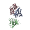
Crystal structure of a putative secreted protein (lpg1979) from Legionella pneumophila subsp. pneumophila str. Philadelphia 1 at 2.65 A resolution
Components
hypothetical protein
Keywords
Structural Genomics / Unknown Function / ORFan protein / new combination of alpha helixes and beta sheets / Joint Center for Structural Genomics / JCSG / Protein Structure Initiative / PSI-BIOLOGY
Function / homology
Single alpha-helices involved in coiled-coils or other helix-helix interfaces - #2710 / Domain of unknown function DUF5624 / Domain of unknown function (DUF5624) / Single alpha-helices involved in coiled-coils or other helix-helix interfaces / Helix non-globular / Special / DI(HYDROXYETHYL)ETHER / TRIETHYLENE GLYCOL / DUF5624 domain-containing protein
Mass: 18.015 Da / Num. of mol.: 257 / Source method: isolated from a natural source / Formula: H2O
-
Details
Has protein modification
Y
Sequence details
THE CONSTRUCT (22-375) WAS EXPRESSED WITH A PURIFICATION TAG MGSDKIHHHHHHENLYFQG. THE TAG WAS ...THE CONSTRUCT (22-375) WAS EXPRESSED WITH A PURIFICATION TAG MGSDKIHHHHHHENLYFQG. THE TAG WAS REMOVED WITH TEV PROTEASE LEAVING ONLY A GLYCINE (0) FOLLOWED BY THE TARGET SEQUENCE.
-
Experimental details
-
Experiment
Experiment
Method: X-RAY DIFFRACTION / Number of used crystals: 1
-
Sample preparation
Crystal
Density Matthews: 2.89 Å3/Da / Density % sol: 57.39 %
Crystal grow
Temperature: 293 K / Method: vapor diffusion, sitting drop / pH: 8.5 Details: 0.2M lithium sulfate, 40.0% polyethylene glycol 400, 0.2M lithium sulfate, 40.0% polyethylene glycol 400, 0.1M TRIS pH 8.5, NANODROP, VAPOR DIFFUSION, SITTING DROP, temperature 293K
Type: DECTRIS PILATUS 6M / Detector: PIXEL / Date: Jan 25, 2013 Details: Flat mirror (vertical focusing); single crystal Si(111) bent monochromator (horizontal focusing)
Radiation
Monochromator: single crystal Si(111) bent / Protocol: MAD / Monochromatic (M) / Laue (L): M / Scattering type: x-ray
Radiation wavelength
ID
Wavelength (Å)
Relative weight
1
0.97932
1
2
0.91837
1
3
0.97858
1
Reflection
Resolution: 2.65→47.97 Å / Num. obs: 33147 / % possible obs: 98.1 % / Observed criterion σ(I): -3 / Biso Wilson estimate: 56.91 Å2 / Rmerge(I) obs: 0.087 / Net I/σ(I): 11.63
Reflection shell
Diffraction-ID: 1
Resolution (Å)
Highest resolution (Å)
Rmerge(I) obs
Mean I/σ(I) obs
Num. measured obs
Num. unique obs
% possible all
2.65-2.74
0.57
2.3
9677
3064
97.5
2.74-2.85
0.419
3
11528
3335
99.7
2.85-2.98
0.304
4.1
11472
3310
99.1
2.98-3.14
0.223
5.5
11550
3354
98.7
3.14-3.34
0.145
8
11370
3373
98.9
3.34-3.59
0.1
10.8
10019
3161
96.6
3.59-3.95
0.071
15.2
11520
3342
99.2
3.95-4.52
0.051
19.7
11471
3355
98.4
4.52-5.67
0.047
20.8
10553
3278
96.4
5.67
0.039
25.3
11382
3554
96.3
-
Phasing
Phasing
Method: MAD
-
Processing
Software
Name
Version
Classification
NB
MolProbity
3beta29
modelbuilding
PDB_EXTRACT
3.1
dataextraction
SHELX
phasing
SHARP
phasing
XSCALE
July4, 2012
datascaling
BUSTER-TNT
2.10.0
refinement
XDS
datareduction
SHELXD
phasing
BUSTER
2.10.0
refinement
Refinement
Method to determine structure: MAD / Resolution: 2.65→47.97 Å / Cor.coef. Fo:Fc: 0.9496 / Cor.coef. Fo:Fc free: 0.9231 / Occupancy max: 1 / Occupancy min: 0.75 / Cross valid method: THROUGHOUT / σ(F): 0 Details: 1. A MET-INHIBITION PROTOCOL WAS USED FOR SELENOMETHIONINE INCORPORATION DURING PROTEIN EXPRESSION. THE OCCUPANCY OF THE SE ATOMS IN THE MSE RESIDUES WAS REDUCED TO 0.75 FOR THE REDUCED ...Details: 1. A MET-INHIBITION PROTOCOL WAS USED FOR SELENOMETHIONINE INCORPORATION DURING PROTEIN EXPRESSION. THE OCCUPANCY OF THE SE ATOMS IN THE MSE RESIDUES WAS REDUCED TO 0.75 FOR THE REDUCED SCATTERING POWER DUE TO PARTIAL S-MET INCORPORATION. 2. ATOM RECORD CONTAINS SUM OF TLS AND RESIDUAL B FACTORS. ANISOU RECORD CONTAINS SUM OF TLS AND RESIDUAL U FACTORS. 3. THE MAD PHASES WERE USED AS RESTRAINTS DURING REFINEMENT. 4. PEG FRAGMENTS, CL AND SO4 MODELED ARE PRESENT IN CRYSTALLIZATION CONDITION. 5.NCS RESTRAINTS WERE IMPOSED BY AUTOBUSTER'S LSSR PROCEDURE (-AUTONCS).
In the structure databanks used in Yorodumi, some data are registered as the other names, "COVID-19 virus" and "2019-nCoV". Here are the details of the virus and the list of structure data.
Jan 31, 2019. EMDB accession codes are about to change! (news from PDBe EMDB page)
EMDB accession codes are about to change! (news from PDBe EMDB page)
The allocation of 4 digits for EMDB accession codes will soon come to an end. Whilst these codes will remain in use, new EMDB accession codes will include an additional digit and will expand incrementally as the available range of codes is exhausted. The current 4-digit format prefixed with “EMD-” (i.e. EMD-XXXX) will advance to a 5-digit format (i.e. EMD-XXXXX), and so on. It is currently estimated that the 4-digit codes will be depleted around Spring 2019, at which point the 5-digit format will come into force.
The EM Navigator/Yorodumi systems omit the EMD- prefix.
Related info.:Q: What is EMD? / ID/Accession-code notation in Yorodumi/EM Navigator
Yorodumi is a browser for structure data from EMDB, PDB, SASBDB, etc.
This page is also the successor to EM Navigator detail page, and also detail information page/front-end page for Omokage search.
The word "yorodu" (or yorozu) is an old Japanese word meaning "ten thousand". "mi" (miru) is to see.
Related info.:EMDB / PDB / SASBDB / Comparison of 3 databanks / Yorodumi Search / Aug 31, 2016. New EM Navigator & Yorodumi / Yorodumi Papers / Jmol/JSmol / Function and homology information / Changes in new EM Navigator and Yorodumi
 Movie
Movie Controller

 Yorodumi
 Open data
 Basic information
 Components
 Keywords
 Function and homology information
 Legionella pneumophila subsp. pneumophila (bacteria)
 X-RAY DIFFRACTION /
  Authors
 Citation
 Structure visualization
 Downloads & links
 Other downloads


 PDBj
PDBj Assembly


 Mass: 35.453 Da / Num. of mol.: 3 / Source method: obtained synthetically / Formula: Cl
 Mass: 96.063 Da / Num. of mol.: 12 / Source method: obtained synthetically / Formula: SO4
 Mass: 150.173 Da / Num. of mol.: 4 / Source method: obtained synthetically / Formula: C6H14O4
 Mass: 238.278 Da / Num. of mol.: 3 / Source method: obtained synthetically / Formula: C10H22O6 / Comment: precipitant*YM
 Mass: 106.120 Da / Num. of mol.: 3 / Source method: obtained synthetically / Formula: C4H10O3
 Sample preparation
 / Beamline: BL11-1 / Wavelength: 0.97932,0.91837,0.97858
 Processing