- PDB-7lir: Structure of the invertebrate ALK GRD -
+
Open data
ID or keywords:
Loading...
-
Basic information
Entry
Database: PDB / ID: 7lir
Title
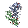
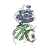
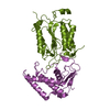
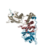
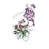
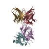
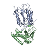
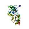
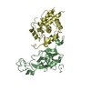
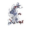
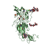
Structure of the invertebrate ALK GRD
Components
ALK tyrosine kinase receptor homolog scd-2
Keywords
SIGNALING PROTEIN / ALK / anaplastic lymphoma kinase / GRD / glycine rich domain / Pole / poly-glycine extension / PXL / poly-glycine extension loops / neuroblastoma / cancer / receptor / kinase / oncogene / AUG / augmentor / ALKAL / ALK and LTK activating ligand / FAM150
Function / homology
Function and homology information
dauer larval development / sensory processing / sensory perception of chemical stimulus / regulation of neuron differentiation / transmembrane receptor protein tyrosine kinase activity / receptor protein-tyrosine kinase / chemotaxis / ATP binding / plasma membrane Similarity search - Function
ALK/LTK, Glycine-rich domain / MAM domain / MAM domain profile. / LDL-receptor class A (LDLRA) domain profile. / Low-density lipoprotein receptor domain class A / Low-density lipoprotein (LDL) receptor class A repeat / : / Tyrosine-protein kinase, catalytic domain / Tyrosine kinase, catalytic domain / Tyrosine protein kinases specific active-site signature. ...ALK/LTK, Glycine-rich domain / MAM domain / MAM domain profile. / LDL-receptor class A (LDLRA) domain profile. / Low-density lipoprotein receptor domain class A / Low-density lipoprotein (LDL) receptor class A repeat / : / Tyrosine-protein kinase, catalytic domain / Tyrosine kinase, catalytic domain / Tyrosine protein kinases specific active-site signature. / Tyrosine-protein kinase, active site / Concanavalin A-like lectin/glucanase domain superfamily / Protein tyrosine and serine/threonine kinase / Serine-threonine/tyrosine-protein kinase, catalytic domain / Protein kinase, ATP binding site / Protein kinases ATP-binding region signature. / Protein kinase domain profile. / Protein kinase domain / Protein kinase-like domain superfamily Similarity search - Domain/homology
Method to determine structure: SAD / Resolution: 2.6→50.18 Å / Cor.coef. Fo:Fc: 0.938 / Cor.coef. Fo:Fc free: 0.908 / SU B: 12.656 / SU ML: 0.247 / SU R Cruickshank DPI: 0.313 / Cross valid method: THROUGHOUT / σ(F): 0 / ESU R: 0.313 / ESU R Free: 0.26 / Stereochemistry target values: MAXIMUM LIKELIHOOD Details: HYDROGENS HAVE BEEN ADDED IN THE RIDING POSITIONS U VALUES : REFINED INDIVIDUALLY
Rfactor
Num. reflection
% reflection
Selection details
Rfree
0.2701
2175
4.8 %
RANDOM
Rwork
0.2233
-
-
-
obs
0.2255
42770
99.99 %
-
Solvent computation
Ion probe radii: 0.8 Å / Shrinkage radii: 0.8 Å / VDW probe radii: 1.2 Å / Solvent model: MASK
In the structure databanks used in Yorodumi, some data are registered as the other names, "COVID-19 virus" and "2019-nCoV". Here are the details of the virus and the list of structure data.
Jan 31, 2019. EMDB accession codes are about to change! (news from PDBe EMDB page)
EMDB accession codes are about to change! (news from PDBe EMDB page)
The allocation of 4 digits for EMDB accession codes will soon come to an end. Whilst these codes will remain in use, new EMDB accession codes will include an additional digit and will expand incrementally as the available range of codes is exhausted. The current 4-digit format prefixed with “EMD-” (i.e. EMD-XXXX) will advance to a 5-digit format (i.e. EMD-XXXXX), and so on. It is currently estimated that the 4-digit codes will be depleted around Spring 2019, at which point the 5-digit format will come into force.
The EM Navigator/Yorodumi systems omit the EMD- prefix.
Related info.:Q: What is EMD? / ID/Accession-code notation in Yorodumi/EM Navigator
Yorodumi is a browser for structure data from EMDB, PDB, SASBDB, etc.
This page is also the successor to EM Navigator detail page, and also detail information page/front-end page for Omokage search.
The word "yorodu" (or yorozu) is an old Japanese word meaning "ten thousand". "mi" (miru) is to see.
Related info.:EMDB / PDB / SASBDB / Comparison of 3 databanks / Yorodumi Search / Aug 31, 2016. New EM Navigator & Yorodumi / Yorodumi Papers / Jmol/JSmol / Function and homology information / Changes in new EM Navigator and Yorodumi
 Movie
Movie Controller


 Open data
 Basic information
 Components
 Keywords
 Function and homology information
 X-RAY DIFFRACTION /
  Authors
 United States, 2items
  Citation
 Structure visualization
 Downloads & links
 Other downloads


 PDBj
PDBj

 Assembly
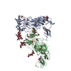
 Trichoplusia ni (cabbage looper)
 Type: D-saccharide, beta linking / Mass: 221.208 Da / Num. of mol.: 1 / Source method: obtained synthetically / Formula: C8H15NO6

 Mass: 96.063 Da / Num. of mol.: 5 / Source method: obtained synthetically / Formula: SO4
 Sample preparation
 Processing