 Movie
Movie Controller

[English] 日本語
 Yorodumi
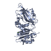
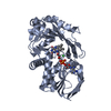
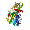
- PDB-7lhk: High-Resolution Crystal Structure of a Lipin/Pah Phosphatidic Aci... -
+ Open data

- Basic information
| Entry | Database: PDB / ID: 7lhk | ||||||||||||
|---|---|---|---|---|---|---|---|---|---|---|---|---|---|
| Title | High-Resolution Crystal Structure of a Lipin/Pah Phosphatidic Acid Phosphatase | ||||||||||||
 Components Components | Nuclear elongation and deformation protein | ||||||||||||
 Keywords Keywords | HYDROLASE / lipin / phosphohydrolase / phosphatidic acid phosphatase / haloalkanoic acid dehalogenase | ||||||||||||
| Function / homology |  Function and homology information Function and homology information | ||||||||||||
| Biological species |   Tetrahymena thermophila (eukaryote) Tetrahymena thermophila (eukaryote) | ||||||||||||
| Method |  X-RAY DIFFRACTION / SYNCHROTRON / MOLECULAR REPLACEMENT / Resolution: 1.95 Å X-RAY DIFFRACTION / SYNCHROTRON / MOLECULAR REPLACEMENT / Resolution: 1.95 Å | ||||||||||||
 Authors Authors | Khayyo, V.I. / Airola, M.V. | ||||||||||||
| Funding support |  United States, 3items United States, 3items
| ||||||||||||
 Citation Citation | Journal: J.Biol.Chem. / Year: 2025 Title: Structures of a lipin/Pah phosphatidic acid phosphatase in distinct catalytic states reveal a signature motif for substrate recognition. Authors: Vitkovska, T. / Welcome, F.S. / Khayyo, V.I. / Gao, S. / Wymore, T. / Airola, M.V. | ||||||||||||
| History |
|
- Structure visualization
| Structure viewer | Molecule: MolmilJmol/JSmol |
|---|
- Downloads & links
-Download
| PDBx/mmCIF format | 7lhk.cif.gz | 308 KB | Display | PDBx/mmCIF format |
|---|---|---|---|---|
| PDB format | pdb7lhk.ent.gz | 202.9 KB | Display | PDB format |
| PDBx/mmJSON format | 7lhk.json.gz | Tree view | PDBx/mmJSON format | |
| Others |  Other downloads Other downloads |
-Validation report
| Arichive directory | https://data.pdbj.org/pub/pdb/validation_reports/lh/7lhkftp://data.pdbj.org/pub/pdb/validation_reports/lh/7lhk | HTTPS FTP |
|---|
-Related structure data
| Related structure data |  9d13C  9d14C  9d15C  9d16C  6tzyS S: Starting model for refinement C: citing same article ( |
|---|---|
| Similar structure data |


-Links
 PDBj
PDBj


- Assembly
| Deposited unit | 
| ||||||||||||
|---|---|---|---|---|---|---|---|---|---|---|---|---|---|
| 1 | 
| ||||||||||||
| 2 | 
| ||||||||||||
| Unit cell |
| ||||||||||||
| Components on special symmetry positions |
|
-Components
| #1: Protein | Mass: 35758.023 Da / Num. of mol.: 2 Source method: isolated from a genetically manipulated source Source: (gene. exp.) Tetrahymena thermophila (strain SB210) (eukaryote)Strain: SB210 / Gene: TTHERM_00215970 / Plasmid: ppSUMO Production host:  References: UniProt: I7MFJ3 #2: Chemical |   Mass: 136.989 Da / Num. of mol.: 2 / Source method: obtained synthetically / Formula: C2H6AsO2 Mass: 136.989 Da / Num. of mol.: 2 / Source method: obtained synthetically / Formula: C2H6AsO2#3: Water | ChemComp-HOH / |  Mass: 18.015 Da / Num. of mol.: 576 / Source method: isolated from a natural source / Formula: H2O Mass: 18.015 Da / Num. of mol.: 576 / Source method: isolated from a natural source / Formula: H2OHas ligand of interest | N | Has protein modification | N | |
|---|
-Experimental details
-Experiment
| Experiment | Method: X-RAY DIFFRACTION / Number of used crystals: 1 |
|---|
- Sample preparation
| Crystal | Density Matthews: 2.72 Å3/Da / Density % sol: 54.82 % |
|---|---|
| Crystal grow | Temperature: 298 K / Method: vapor diffusion, hanging drop / pH: 6.5 Details: Tt Pah2 in a ratio of 2:1 with well solution of 0.1M sodium cacodylate pH 6.5, 1 M NaCl, 10% glycerol, 30% PEG 600 |
-Data collection
| Diffraction | Mean temperature: 100 K / Serial crystal experiment: N |
|---|---|
| Diffraction source | Source: SYNCHROTRON / Site: APS / Beamline: 23-ID-B / Wavelength: 0.97934 Å |
| Detector | Type: DECTRIS EIGER X 16M / Detector: PIXEL / Date: Jul 23, 2020 |
| Radiation | Protocol: SINGLE WAVELENGTH / Monochromatic (M) / Laue (L): M / Scattering type: x-ray |
| Radiation wavelength | Wavelength: 0.97934 Å / Relative weight: 1 |
| Reflection | Resolution: 1.95→55.25 Å / Num. obs: 720604 / % possible obs: 99.54 % / Redundancy: 12.6 % / Biso Wilson estimate: 30.57 Å2 / CC1/2: 0.999 / CC star: 1 / Rmerge(I) obs: 0.08466 / Rpim(I) all: 0.02437 / Rrim(I) all: 0.08818 / Net I/σ(I): 16.77 |
| Reflection shell | Resolution: 1.95→2.02 Å / Redundancy: 7.9 % / Rmerge(I) obs: 0.686 / Mean I/σ(I) obs: 2.87 / Num. unique obs: 5410 / CC1/2: 0.907 / CC star: 0.975 / Rpim(I) all: 0.2381 / Rrim(I) all: 0.686 / % possible all: 95.83 |
- Processing
| Software |
| |||||||||||||||||||||||||||||||||||||||||||||||||||||||||||||||||||||||||||||||||||||||||||||||||||||||||||||||||||||||||||||||||||||||||||||||||||
|---|---|---|---|---|---|---|---|---|---|---|---|---|---|---|---|---|---|---|---|---|---|---|---|---|---|---|---|---|---|---|---|---|---|---|---|---|---|---|---|---|---|---|---|---|---|---|---|---|---|---|---|---|---|---|---|---|---|---|---|---|---|---|---|---|---|---|---|---|---|---|---|---|---|---|---|---|---|---|---|---|---|---|---|---|---|---|---|---|---|---|---|---|---|---|---|---|---|---|---|---|---|---|---|---|---|---|---|---|---|---|---|---|---|---|---|---|---|---|---|---|---|---|---|---|---|---|---|---|---|---|---|---|---|---|---|---|---|---|---|---|---|---|---|---|---|---|---|---|
| Refinement | Method to determine structure: MOLECULAR REPLACEMENT Starting model: 6TZY Resolution: 1.95→55.25 Å / SU ML: 0.1972 / Cross valid method: FREE R-VALUE / σ(F): 0 / Phase error: 20.6774 / Stereochemistry target values: CDL v1.2
| |||||||||||||||||||||||||||||||||||||||||||||||||||||||||||||||||||||||||||||||||||||||||||||||||||||||||||||||||||||||||||||||||||||||||||||||||||
| Solvent computation | Shrinkage radii: 0.9 Å / VDW probe radii: 1.11 Å / Solvent model: FLAT BULK SOLVENT MODEL | |||||||||||||||||||||||||||||||||||||||||||||||||||||||||||||||||||||||||||||||||||||||||||||||||||||||||||||||||||||||||||||||||||||||||||||||||||
| Displacement parameters | Biso mean: 27.45 Å2 | |||||||||||||||||||||||||||||||||||||||||||||||||||||||||||||||||||||||||||||||||||||||||||||||||||||||||||||||||||||||||||||||||||||||||||||||||||
| Refinement step | Cycle: LAST / Resolution: 1.95→55.25 Å
| |||||||||||||||||||||||||||||||||||||||||||||||||||||||||||||||||||||||||||||||||||||||||||||||||||||||||||||||||||||||||||||||||||||||||||||||||||
| Refine LS restraints |
| |||||||||||||||||||||||||||||||||||||||||||||||||||||||||||||||||||||||||||||||||||||||||||||||||||||||||||||||||||||||||||||||||||||||||||||||||||
| LS refinement shell |
|
