 Movie
Movie Controller


+ Open data

- Basic information
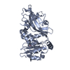
| Entry | Database: PDB / ID: 9d13 | ||||||
|---|---|---|---|---|---|---|---|
| Title | Tt Pah2 D148N delta helix apo | ||||||
 Components Components | Nuclear elongation and deformation protein | ||||||
 Keywords Keywords | HYDROLASE / lipin Pah phosphatidic acid phosphatase / lipid phosphatase | ||||||
| Function / homology |  Function and homology information Function and homology information | ||||||
| Biological species |   Tetrahymena thermophila (eukaryote) Tetrahymena thermophila (eukaryote) | ||||||
| Method |  X-RAY DIFFRACTION / SYNCHROTRON / MOLECULAR REPLACEMENT / Resolution: 1.98 Å X-RAY DIFFRACTION / SYNCHROTRON / MOLECULAR REPLACEMENT / Resolution: 1.98 Å | ||||||
 Authors Authors | Vitkovska, T. / Khayyo, V.I. / Airola, M.V. | ||||||
| Funding support |  United States, 1items United States, 1items
| ||||||
 Citation Citation | Journal: J.Biol.Chem. / Year: 2025 Title: Structures of a lipin/Pah phosphatidic acid phosphatase in distinct catalytic states reveal a signature motif for substrate recognition. Authors: Vitkovska, T. / Welcome, F.S. / Khayyo, V.I. / Gao, S. / Wymore, T. / Airola, M.V. | ||||||
| History |
|
- Structure visualization
| Structure viewer | Molecule: MolmilJmol/JSmol |
|---|
- Downloads & links
-Download
| PDBx/mmCIF format | 9d13.cif.gz | 215.8 KB | Display | PDBx/mmCIF format |
|---|---|---|---|---|
| PDB format | pdb9d13.ent.gz | 146.2 KB | Display | PDB format |
| PDBx/mmJSON format | 9d13.json.gz | Tree view | PDBx/mmJSON format | |
| Others |  Other downloads Other downloads |
-Validation report
| Arichive directory | https://data.pdbj.org/pub/pdb/validation_reports/d1/9d13ftp://data.pdbj.org/pub/pdb/validation_reports/d1/9d13 | HTTPS FTP |
|---|
-Related structure data


-Links
 PDBj
PDBj- Assembly
| Deposited unit | 
| ||||||||||||
|---|---|---|---|---|---|---|---|---|---|---|---|---|---|
| 1 |
| ||||||||||||
| Unit cell |
|
-Components
| #1: Protein | Mass: 34479.613 Da / Num. of mol.: 1 / Mutation: D148N Source method: isolated from a genetically manipulated source Details: Tt Pah2 residues 21-321, D148N / Source: (gene. exp.) Tetrahymena thermophila (eukaryote) / Gene: TTHERM_00215970 / Production host:  |
|---|---|
| #2: Chemical | ChemComp-GOL /   Mass: 92.094 Da / Num. of mol.: 1 / Source method: obtained synthetically / Formula: C3H8O3 Mass: 92.094 Da / Num. of mol.: 1 / Source method: obtained synthetically / Formula: C3H8O3 |
| #3: Water | ChemComp-HOH /  Mass: 18.015 Da / Num. of mol.: 104 / Source method: isolated from a natural source / Formula: H2O Mass: 18.015 Da / Num. of mol.: 104 / Source method: isolated from a natural source / Formula: H2O |
| Has ligand of interest | N |
| Has protein modification | N |
-Experimental details
-Experiment
| Experiment | Method: X-RAY DIFFRACTION / Number of used crystals: 1 |
|---|
- Sample preparation
| Crystal | Density Matthews: 2.14 Å3/Da / Density % sol: 42.41 % |
|---|---|
| Crystal grow | Temperature: 298 K / Method: vapor diffusion, hanging drop / Details: 30% PEG 3000, 0.1 M CHES pH 8.5 - 9.5 |
-Data collection
| Diffraction | Mean temperature: 100 K / Serial crystal experiment: N |
|---|---|
| Diffraction source | Source: SYNCHROTRON / Site: APS / Beamline: 23-ID-D / Wavelength: 1.0332 Å |
| Detector | Type: DECTRIS PILATUS 6M / Detector: PIXEL / Date: Jun 3, 2021 |
| Radiation | Protocol: SINGLE WAVELENGTH / Monochromatic (M) / Laue (L): M / Scattering type: x-ray |
| Radiation wavelength | Wavelength: 1.0332 Å / Relative weight: 1 |
| Reflection | Resolution: 1.98→60.78 Å / Num. obs: 21672 / % possible obs: 99.79 % / Redundancy: 12.2 % / Biso Wilson estimate: 40.41 Å2 / CC1/2: 0.999 / CC star: 1 / Rmerge(I) obs: 0.09526 / Rpim(I) all: 0.02848 / Rrim(I) all: 0.09957 / Net I/σ(I): 14.78 |
| Reflection shell | Resolution: 1.98→2.05 Å / Redundancy: 12.3 % / Rmerge(I) obs: 2.157 / Mean I/σ(I) obs: 1.58 / Num. unique obs: 2110 / CC1/2: 0.67 / CC star: 0.896 / Rpim(I) all: 0.6363 / Rrim(I) all: 2.251 / % possible all: 99.39 |
- Processing
| Software |
| |||||||||||||||||||||||||||||||||||||||||||||||||||||||||||||||||||||||||||||||||||||||||||||||||||||||||
|---|---|---|---|---|---|---|---|---|---|---|---|---|---|---|---|---|---|---|---|---|---|---|---|---|---|---|---|---|---|---|---|---|---|---|---|---|---|---|---|---|---|---|---|---|---|---|---|---|---|---|---|---|---|---|---|---|---|---|---|---|---|---|---|---|---|---|---|---|---|---|---|---|---|---|---|---|---|---|---|---|---|---|---|---|---|---|---|---|---|---|---|---|---|---|---|---|---|---|---|---|---|---|---|---|---|---|
| Refinement | Method to determine structure: MOLECULAR REPLACEMENT / Resolution: 1.98→60.78 Å / SU ML: 0.2386 / Cross valid method: FREE R-VALUE / σ(F): 1.34 / Phase error: 24.9454 Stereochemistry target values: GeoStd + Monomer Library + CDL v1.2
| |||||||||||||||||||||||||||||||||||||||||||||||||||||||||||||||||||||||||||||||||||||||||||||||||||||||||
| Solvent computation | Shrinkage radii: 0.9 Å / VDW probe radii: 1.1 Å / Solvent model: FLAT BULK SOLVENT MODEL | |||||||||||||||||||||||||||||||||||||||||||||||||||||||||||||||||||||||||||||||||||||||||||||||||||||||||
| Displacement parameters | Biso mean: 52.77 Å2 | |||||||||||||||||||||||||||||||||||||||||||||||||||||||||||||||||||||||||||||||||||||||||||||||||||||||||
| Refinement step | Cycle: LAST / Resolution: 1.98→60.78 Å
| |||||||||||||||||||||||||||||||||||||||||||||||||||||||||||||||||||||||||||||||||||||||||||||||||||||||||
| Refine LS restraints |
| |||||||||||||||||||||||||||||||||||||||||||||||||||||||||||||||||||||||||||||||||||||||||||||||||||||||||
| LS refinement shell |
| |||||||||||||||||||||||||||||||||||||||||||||||||||||||||||||||||||||||||||||||||||||||||||||||||||||||||
| Refinement TLS params. | Method: refined / Origin x: 21.7870815321 Å / Origin y: 7.75432270096 Å / Origin z: -18.4079615349 Å
| |||||||||||||||||||||||||||||||||||||||||||||||||||||||||||||||||||||||||||||||||||||||||||||||||||||||||
| Refinement TLS group | Selection details: all |
