 Movie
Movie Controller

[English] 日本語
 Yorodumi
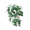
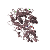
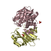
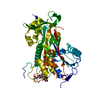
- PDB-7lcu: X-ray structure of Furin bound to BOS-318, a small molecule inhibitor -
+ Open data

- Basic information
| Entry | Database: PDB / ID: 7lcu | ||||||
|---|---|---|---|---|---|---|---|
| Title | X-ray structure of Furin bound to BOS-318, a small molecule inhibitor | ||||||
 Components Components | Furin | ||||||
 Keywords Keywords | HYDROLASE/INHIBITOR / Inhibitor protease / HYDROLASE-INHIBITOR complex | ||||||
| Function / homology |  Function and homology information Function and homology informationfurin / nerve growth factor production / dibasic protein processing / plasma lipoprotein particle remodeling / NGF processing / viral translation / negative regulation of transforming growth factor beta1 production / Assembly of active LPL and LIPC lipase complexes / regulation of cholesterol transport / : ...furin / nerve growth factor production / dibasic protein processing / plasma lipoprotein particle remodeling / NGF processing / viral translation / negative regulation of transforming growth factor beta1 production / Assembly of active LPL and LIPC lipase complexes / regulation of cholesterol transport / : / negative regulation of low-density lipoprotein particle receptor catabolic process / peptide biosynthetic process / Pre-NOTCH Processing in Golgi / nerve growth factor binding / Synthesis and processing of ENV and VPU / cytokine precursor processing / Formation of the cornified envelope / secretion by cell / Signaling by PDGF / trans-Golgi network transport vesicle / Signaling by NODAL / heparan sulfate binding / Elastic fibre formation / blastocyst formation / positive regulation of membrane protein ectodomain proteolysis / peptide hormone processing / zymogen activation / CD163 mediating an anti-inflammatory response / Activation of Matrix Metalloproteinases / regulation of protein catabolic process / Collagen degradation / collagen catabolic process / TGF-beta receptor signaling activates SMADs / Maturation of hRSV A proteins / Respiratory syncytial virus (RSV) attachment and entry / Uptake and function of anthrax toxins / extracellular matrix disassembly / regulation of signal transduction / endopeptidase activator activity / Removal of aminoterminal propeptides from gamma-carboxylated proteins / viral life cycle / extracellular matrix organization / peptide binding / transforming growth factor beta receptor signaling pathway / serine-type peptidase activity / negative regulation of inflammatory response to antigenic stimulus / serine-type endopeptidase inhibitor activity / protein maturation / trans-Golgi network / SMAD2/SMAD3:SMAD4 heterotrimer regulates transcription / protein processing / Golgi lumen / peptidase activity / heparin binding / protease binding / endopeptidase activity / amyloid fibril formation / Induction of Cell-Cell Fusion / Potential therapeutics for SARS / positive regulation of viral entry into host cell / Attachment and Entry / endosome membrane / viral protein processing / membrane raft / Amyloid fiber formation / Golgi membrane / serine-type endopeptidase activity / cell surface / endoplasmic reticulum / extracellular exosome / extracellular region / membrane / metal ion binding / plasma membrane Similarity search - Function | ||||||
| Biological species |  Homo sapiens (human) Homo sapiens (human) | ||||||
| Method |  X-RAY DIFFRACTION / SYNCHROTRON / MOLECULAR REPLACEMENT / Resolution: 1.24 Å X-RAY DIFFRACTION / SYNCHROTRON / MOLECULAR REPLACEMENT / Resolution: 1.24 Å | ||||||
 Authors Authors | Campobasso, N. / Reid, R. | ||||||
 Citation Citation | Journal: Cell Chem Biol / Year: 2022 Title: A highly selective, cell-permeable furin inhibitor BOS-318 rescues key features of cystic fibrosis airway disease. Authors: Douglas, L.E.J. / Reihill, J.A. / Ho, M.W.Y. / Axten, J.M. / Campobasso, N. / Schneck, J.L. / Rendina, A.R. / Wilcoxen, K.M. / Martin, S.L. #1: Journal: Acta Crystallogr.,Sect.F / Year: 2019Title: BacMam production and crystal structure of nonglycosylated apo human furin at 1.89 angstrom resolution. Authors: Pearce, K.H. / Overton, L.K. / Gampe, R.T. / Barrett, G.B. / Taylor, J.D. / McKee, D.D. / Campobasso, N. / Nolte, R.T. / Reid, R.A. | ||||||
| History |
|
- Structure visualization
| Structure viewer | Molecule: MolmilJmol/JSmol |
|---|
- Downloads & links
-Download
| PDBx/mmCIF format | 7lcu.cif.gz | 145 KB | Display | PDBx/mmCIF format |
|---|---|---|---|---|
| PDB format | pdb7lcu.ent.gz | 89 KB | Display | PDB format |
| PDBx/mmJSON format | 7lcu.json.gz | Tree view | PDBx/mmJSON format | |
| Others |  Other downloads Other downloads |
-Validation report
| Arichive directory | https://data.pdbj.org/pub/pdb/validation_reports/lc/7lcuftp://data.pdbj.org/pub/pdb/validation_reports/lc/7lcu | HTTPS FTP |
|---|
-Related structure data
| Related structure data |  4z2aS S: Starting model for refinement |
|---|---|
| Similar structure data |


-Links
 PDBj
PDBj


- Assembly
| Deposited unit | 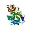
| ||||||||||||
|---|---|---|---|---|---|---|---|---|---|---|---|---|---|
| 1 |
| ||||||||||||
| Unit cell |
| ||||||||||||
| Components on special symmetry positions |
|
-Components
| #1: Protein | Mass: 53511.602 Da / Num. of mol.: 1 / Fragment: UNP residues 108-574 Source method: isolated from a genetically manipulated source Source: (gene. exp.) Homo sapiens (human) / Gene: FURIN, FUR, PACE, PCSK3 / Production host:  Insect BA phytoplasma (bacteria) / References: UniProt: P09958, furin Insect BA phytoplasma (bacteria) / References: UniProt: P09958, furin | ||||||||||
|---|---|---|---|---|---|---|---|---|---|---|---|
| #2: Chemical | ChemComp-CA /   Mass: 40.078 Da / Num. of mol.: 4 / Source method: obtained synthetically / Formula: Ca Mass: 40.078 Da / Num. of mol.: 4 / Source method: obtained synthetically / Formula: Ca#3: Chemical | ChemComp-XTA / ( |   Mass: 571.498 Da / Num. of mol.: 1 / Source method: obtained synthetically / Formula: C28H32Cl2N6O3 / Feature type: SUBJECT OF INVESTIGATION Mass: 571.498 Da / Num. of mol.: 1 / Source method: obtained synthetically / Formula: C28H32Cl2N6O3 / Feature type: SUBJECT OF INVESTIGATION#4: Chemical |   Mass: 62.068 Da / Num. of mol.: 2 / Source method: obtained synthetically / Formula: C2H6O2 Mass: 62.068 Da / Num. of mol.: 2 / Source method: obtained synthetically / Formula: C2H6O2#5: Water | ChemComp-HOH / |  Mass: 18.015 Da / Num. of mol.: 352 / Source method: isolated from a natural source / Formula: H2O Mass: 18.015 Da / Num. of mol.: 352 / Source method: isolated from a natural source / Formula: H2OHas ligand of interest | Y | Has protein modification | Y | |
-Experimental details
-Experiment
| Experiment | Method: X-RAY DIFFRACTION / Number of used crystals: 1 |
|---|
- Sample preparation
| Crystal | Density Matthews: 2.25 Å3/Da / Density % sol: 45.43 % |
|---|---|
| Crystal grow | Temperature: 277 K / Method: vapor diffusion / pH: 7.5 / Details: PEG8000, potassium dihydrogen phosphate |
-Data collection
| Diffraction | Mean temperature: 100 K / Serial crystal experiment: N |
|---|---|
| Diffraction source | Source: SYNCHROTRON / Site: APS  / Beamline: 21-ID-G / Wavelength: 0.9785 Å / Beamline: 21-ID-G / Wavelength: 0.9785 Å |
| Detector | Type: DECTRIS PILATUS 300K / Detector: PIXEL / Date: Mar 4, 2016 |
| Radiation | Monochromator: diamond(111) / Protocol: SINGLE WAVELENGTH / Monochromatic (M) / Laue (L): M / Scattering type: x-ray |
| Radiation wavelength | Wavelength: 0.9785 Å / Relative weight: 1 |
| Reflection | Resolution: 1.24→48 Å / Num. obs: 131144 / % possible obs: 96 % / Redundancy: 2 % / Biso Wilson estimate: 10.3 Å2 / CC1/2: 0.99 / Net I/σ(I): 15.6 |
| Reflection shell | Resolution: 1.24→1.28 Å / Num. unique obs: 13009 / CC1/2: 0.825 / % possible all: 96 |
- Processing
| Software |
| |||||||||||||||||||||||||||||||||||||||||||||||||||||||||||||||||||||||||||||||||||||||||||||||||||||||||||||||||||||||||||||||||||||||||||||||||||||||||||||||||||||||||||||||||||||||||||||||||||||||||||||||||||||||||
|---|---|---|---|---|---|---|---|---|---|---|---|---|---|---|---|---|---|---|---|---|---|---|---|---|---|---|---|---|---|---|---|---|---|---|---|---|---|---|---|---|---|---|---|---|---|---|---|---|---|---|---|---|---|---|---|---|---|---|---|---|---|---|---|---|---|---|---|---|---|---|---|---|---|---|---|---|---|---|---|---|---|---|---|---|---|---|---|---|---|---|---|---|---|---|---|---|---|---|---|---|---|---|---|---|---|---|---|---|---|---|---|---|---|---|---|---|---|---|---|---|---|---|---|---|---|---|---|---|---|---|---|---|---|---|---|---|---|---|---|---|---|---|---|---|---|---|---|---|---|---|---|---|---|---|---|---|---|---|---|---|---|---|---|---|---|---|---|---|---|---|---|---|---|---|---|---|---|---|---|---|---|---|---|---|---|---|---|---|---|---|---|---|---|---|---|---|---|---|---|---|---|---|---|---|---|---|---|---|---|---|---|---|---|---|---|---|---|---|
| Refinement | Method to determine structure: MOLECULAR REPLACEMENT Starting model: PDB entry 4Z2A Resolution: 1.24→48 Å / SU ML: 0.1103 / Cross valid method: FREE R-VALUE / σ(F): 1.35 / Phase error: 19.8426 Stereochemistry target values: GeoStd + Monomer Library + CDL v1.2 + omega-cdl
| |||||||||||||||||||||||||||||||||||||||||||||||||||||||||||||||||||||||||||||||||||||||||||||||||||||||||||||||||||||||||||||||||||||||||||||||||||||||||||||||||||||||||||||||||||||||||||||||||||||||||||||||||||||||||
| Solvent computation | Shrinkage radii: 0.9 Å / VDW probe radii: 1.11 Å / Solvent model: FLAT BULK SOLVENT MODEL | |||||||||||||||||||||||||||||||||||||||||||||||||||||||||||||||||||||||||||||||||||||||||||||||||||||||||||||||||||||||||||||||||||||||||||||||||||||||||||||||||||||||||||||||||||||||||||||||||||||||||||||||||||||||||
| Displacement parameters | Biso mean: 14.5 Å2 | |||||||||||||||||||||||||||||||||||||||||||||||||||||||||||||||||||||||||||||||||||||||||||||||||||||||||||||||||||||||||||||||||||||||||||||||||||||||||||||||||||||||||||||||||||||||||||||||||||||||||||||||||||||||||
| Refinement step | Cycle: LAST / Resolution: 1.24→48 Å
| |||||||||||||||||||||||||||||||||||||||||||||||||||||||||||||||||||||||||||||||||||||||||||||||||||||||||||||||||||||||||||||||||||||||||||||||||||||||||||||||||||||||||||||||||||||||||||||||||||||||||||||||||||||||||
| Refine LS restraints |
| |||||||||||||||||||||||||||||||||||||||||||||||||||||||||||||||||||||||||||||||||||||||||||||||||||||||||||||||||||||||||||||||||||||||||||||||||||||||||||||||||||||||||||||||||||||||||||||||||||||||||||||||||||||||||
| LS refinement shell |
|
