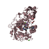
 Movie
Movie Controller


+ Open data

- Basic information
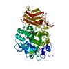
| Entry | Database: PDB / ID: 4z2a | ||||||
|---|---|---|---|---|---|---|---|
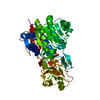
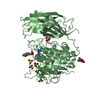
| Title | Crystal structure of unglycosylated apo human furin @1.89A | ||||||
 Components Components | Furin | ||||||
 Keywords Keywords | HYDROLASE / unglycosylated / apo / serine proteinase | ||||||
| Function / homology |  Function and homology information Function and homology informationfurin / nerve growth factor production / dibasic protein processing / plasma lipoprotein particle remodeling / NGF processing / viral translation / negative regulation of transforming growth factor beta1 production / Assembly of active LPL and LIPC lipase complexes / regulation of cholesterol transport / : ...furin / nerve growth factor production / dibasic protein processing / plasma lipoprotein particle remodeling / NGF processing / viral translation / negative regulation of transforming growth factor beta1 production / Assembly of active LPL and LIPC lipase complexes / regulation of cholesterol transport / : / negative regulation of low-density lipoprotein particle receptor catabolic process / peptide biosynthetic process / Pre-NOTCH Processing in Golgi / nerve growth factor binding / Synthesis and processing of ENV and VPU / cytokine precursor processing / Formation of the cornified envelope / secretion by cell / Signaling by PDGF / trans-Golgi network transport vesicle / Signaling by NODAL / heparan sulfate binding / Elastic fibre formation / blastocyst formation / positive regulation of membrane protein ectodomain proteolysis / peptide hormone processing / zymogen activation / CD163 mediating an anti-inflammatory response / Activation of Matrix Metalloproteinases / regulation of protein catabolic process / Collagen degradation / collagen catabolic process / TGF-beta receptor signaling activates SMADs / Maturation of hRSV A proteins / Respiratory syncytial virus (RSV) attachment and entry / Uptake and function of anthrax toxins / extracellular matrix disassembly / regulation of signal transduction / endopeptidase activator activity / Removal of aminoterminal propeptides from gamma-carboxylated proteins / viral life cycle / extracellular matrix organization / peptide binding / transforming growth factor beta receptor signaling pathway / serine-type peptidase activity / negative regulation of inflammatory response to antigenic stimulus / serine-type endopeptidase inhibitor activity / protein maturation / trans-Golgi network / SMAD2/SMAD3:SMAD4 heterotrimer regulates transcription / protein processing / Golgi lumen / peptidase activity / heparin binding / protease binding / endopeptidase activity / amyloid fibril formation / Induction of Cell-Cell Fusion / Potential therapeutics for SARS / positive regulation of viral entry into host cell / Attachment and Entry / endosome membrane / viral protein processing / membrane raft / Amyloid fiber formation / Golgi membrane / serine-type endopeptidase activity / cell surface / endoplasmic reticulum / extracellular exosome / extracellular region / membrane / metal ion binding / plasma membrane Similarity search - Function | ||||||
| Biological species |  Homo sapiens (human) Homo sapiens (human) | ||||||
| Method |  X-RAY DIFFRACTION / MOLECULAR REPLACEMENT / Resolution: 1.89 Å X-RAY DIFFRACTION / MOLECULAR REPLACEMENT / Resolution: 1.89 Å | ||||||
 Authors Authors | Gampe, R.T. / Pearce, K. / Reid, R. | ||||||
 Citation Citation | Journal: Acta Crystallogr.,Sect.F / Year: 2019 Title: BacMam production and crystal structure of nonglycosylated apo human furin at 1.89 angstrom resolution. Authors: Pearce, K.H. / Overton, L.K. / Gampe, R.T. / Barrett, G.B. / Taylor, J.D. / McKee, D.D. / Campobasso, N. / Nolte, R.T. / Reid, R.A. #1: Journal: Acta Crystallogr.,Sect.F / Year: 2019Title: BacMam production and crystal structure of nonglycosylated apo human furin at 1.89A resolution Authors: Pearce, K.H. / Overton, L.K. / Grampe, R.T. / Barret, G.B. / Taylor, J.D. / McKee, D.D. / Campobassom, N. / Nolte, R.T. / Reid, R.A. | ||||||
| History |
|
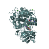
- Structure visualization
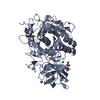
| Structure viewer | Molecule: MolmilJmol/JSmol |
|---|
- Downloads & links
-Download
| PDBx/mmCIF format | 4z2a.cif.gz | 116.6 KB | Display | PDBx/mmCIF format |
|---|---|---|---|---|
| PDB format | pdb4z2a.ent.gz | 85.1 KB | Display | PDB format |
| PDBx/mmJSON format | 4z2a.json.gz | Tree view | PDBx/mmJSON format | |
| Others |  Other downloads Other downloads |
-Validation report
| Arichive directory | https://data.pdbj.org/pub/pdb/validation_reports/z2/4z2aftp://data.pdbj.org/pub/pdb/validation_reports/z2/4z2a | HTTPS FTP |
|---|
-Related structure data
| Related structure data |  1p8jS S: Starting model for refinement |
|---|---|
| Similar structure data |


-Links
 PDBj
PDBj


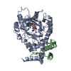
- Assembly
| Deposited unit | 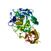
| ||||||||
|---|---|---|---|---|---|---|---|---|---|
| 1 |
| ||||||||
| Unit cell |
|
-Components
| #1: Protein | Mass: 50505.551 Da / Num. of mol.: 1 / Fragment: UNP Residues 110-74 / Mutation: N387D, N440D Source method: isolated from a genetically manipulated source Source: (gene. exp.) Homo sapiens (human) / Gene: FURIN, FUR, PACE, PCSK3 / Production host:   Cricetulus griseus (Chinese hamster) / References: UniProt: P09958, furin Cricetulus griseus (Chinese hamster) / References: UniProt: P09958, furin | ||||||||
|---|---|---|---|---|---|---|---|---|---|
| #2: Chemical | ChemComp-CA /   Mass: 40.078 Da / Num. of mol.: 4 / Source method: obtained synthetically / Formula: Ca Mass: 40.078 Da / Num. of mol.: 4 / Source method: obtained synthetically / Formula: Ca#3: Chemical |   Mass: 94.971 Da / Num. of mol.: 2 / Source method: obtained synthetically / Formula: PO4 Mass: 94.971 Da / Num. of mol.: 2 / Source method: obtained synthetically / Formula: PO4#4: Chemical | ChemComp-EDO / |   Mass: 62.068 Da / Num. of mol.: 1 / Source method: obtained synthetically / Formula: C2H6O2 Mass: 62.068 Da / Num. of mol.: 1 / Source method: obtained synthetically / Formula: C2H6O2#5: Water | ChemComp-HOH / |  Mass: 18.015 Da / Num. of mol.: 408 / Source method: isolated from a natural source / Formula: H2O Mass: 18.015 Da / Num. of mol.: 408 / Source method: isolated from a natural source / Formula: H2OHas protein modification | Y | |
-Experimental details
-Experiment
| Experiment | Method: X-RAY DIFFRACTION / Number of used crystals: 1 |
|---|
- Sample preparation
| Crystal | Density Matthews: 2.36 Å3/Da / Density % sol: 47.94 % |
|---|---|
| Crystal grow | Temperature: 277 K / Method: vapor diffusion, sitting drop Details: Well 13% PEG 8K, 0.110M K-dihydrogen phosphate, 0.1M HEPES pH 7.5, 350 uL protein with 350uL well drops, MRC sitting drop |
-Data collection
| Diffraction | Mean temperature: 100 K | ||||||||||||||||||||||||||||||||||||||||||||||||||||||||||||||||||
|---|---|---|---|---|---|---|---|---|---|---|---|---|---|---|---|---|---|---|---|---|---|---|---|---|---|---|---|---|---|---|---|---|---|---|---|---|---|---|---|---|---|---|---|---|---|---|---|---|---|---|---|---|---|---|---|---|---|---|---|---|---|---|---|---|---|---|---|
| Diffraction source | Source: ROTATING ANODE / Type: RIGAKU FR-E SUPERBRIGHT / Wavelength: 1.54 Å | ||||||||||||||||||||||||||||||||||||||||||||||||||||||||||||||||||
| Detector | Type: RIGAKU SATURN 944+ / Detector: CCD / Date: Aug 10, 2012 | ||||||||||||||||||||||||||||||||||||||||||||||||||||||||||||||||||
| Radiation | Protocol: SINGLE WAVELENGTH / Monochromatic (M) / Laue (L): M / Scattering type: x-ray | ||||||||||||||||||||||||||||||||||||||||||||||||||||||||||||||||||
| Radiation wavelength | Wavelength: 1.54 Å / Relative weight: 1 | ||||||||||||||||||||||||||||||||||||||||||||||||||||||||||||||||||
| Reflection | Resolution: 1.89→50 Å / Num. obs: 37707 / % possible obs: 99.8 % / Redundancy: 3.6 % / Biso Wilson estimate: 11.83 Å2 / Rmerge(I) obs: 0.049 / Χ2: 1.006 / Net I/av σ(I): 25.769 / Net I/σ(I): 13.7 / Num. measured all: 135597 | ||||||||||||||||||||||||||||||||||||||||||||||||||||||||||||||||||
| Reflection shell | Diffraction-ID: 1 / Rejects: _
|
- Processing
| Software |
| |||||||||||||||||||||||||||||||||||||||||||||||||||||||||||||||
|---|---|---|---|---|---|---|---|---|---|---|---|---|---|---|---|---|---|---|---|---|---|---|---|---|---|---|---|---|---|---|---|---|---|---|---|---|---|---|---|---|---|---|---|---|---|---|---|---|---|---|---|---|---|---|---|---|---|---|---|---|---|---|---|---|
| Refinement | Method to determine structure: MOLECULAR REPLACEMENT Starting model: 1P8J Resolution: 1.89→29.581 Å / SU ML: 0.15 / Cross valid method: FREE R-VALUE / σ(F): 1.34 / Phase error: 18.38 / Stereochemistry target values: ML
| |||||||||||||||||||||||||||||||||||||||||||||||||||||||||||||||
| Solvent computation | Shrinkage radii: 0.9 Å / VDW probe radii: 1.11 Å / Solvent model: FLAT BULK SOLVENT MODEL | |||||||||||||||||||||||||||||||||||||||||||||||||||||||||||||||
| Displacement parameters | Biso max: 57.78 Å2 / Biso mean: 16.9953 Å2 / Biso min: 3.04 Å2 | |||||||||||||||||||||||||||||||||||||||||||||||||||||||||||||||
| Refinement step | Cycle: final / Resolution: 1.89→29.581 Å
| |||||||||||||||||||||||||||||||||||||||||||||||||||||||||||||||
| Refine LS restraints |
| |||||||||||||||||||||||||||||||||||||||||||||||||||||||||||||||
| LS refinement shell | Refine-ID: X-RAY DIFFRACTION / Total num. of bins used: 8
|
