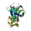
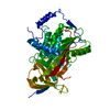
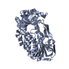
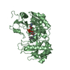
[English] 日本語
Yorodumi
- PDB-5m3y: Crystal structure of human glycosylated angiotensinogen -

+
Open data


ID or keywords:

Loading...

-
Basic information

Entry
Database: PDB / ID: 5m3y
TitleCrystal structure of human glycosylated angiotensinogen
ComponentsAngiotensinogenAngiotensin
KeywordsSerine protease inhibitor / glycosylated / human angiotensiogen
Function / homology
Function and homology information


regulation of blood volume by renin-angiotensin / response to muscle activity involved in regulation of muscle adaptation / regulation of renal sodium excretion / : / type 2 angiotensin receptor binding / maintenance of blood vessel diameter homeostasis by renin-angiotensin / negative regulation of neurotrophin TRK receptor signaling pathway / regulation of extracellular matrix assembly / positive regulation of activation of Janus kinase activity / regulation of renal output by angiotensin ...regulation of blood volume by renin-angiotensin / response to muscle activity involved in regulation of muscle adaptation / regulation of renal sodium excretion / : / type 2 angiotensin receptor binding / maintenance of blood vessel diameter homeostasis by renin-angiotensin / negative regulation of neurotrophin TRK receptor signaling pathway / regulation of extracellular matrix assembly / positive regulation of activation of Janus kinase activity / regulation of renal output by angiotensin / positive regulation of NAD(P)H oxidase activity / G protein-coupled receptor signaling pathway coupled to cGMP nucleotide second messenger / renin-angiotensin regulation of aldosterone production / renal system process / positive regulation of branching involved in ureteric bud morphogenesis / positive regulation of CoA-transferase activity / positive regulation of extracellular matrix assembly / positive regulation of macrophage derived foam cell differentiation / low-density lipoprotein particle remodeling / vasoconstriction / type 1 angiotensin receptor binding / response to angiotensin / positive regulation of extrinsic apoptotic signaling pathway / positive regulation of epidermal growth factor receptor signaling pathway / positive regulation of cardiac muscle hypertrophy / positive regulation of gap junction assembly / regulation of vasoconstriction / regulation of cardiac conduction / positive regulation of epithelial to mesenchymal transition / blood vessel remodeling / positive regulation of protein tyrosine kinase activity / Metabolism of Angiotensinogen to Angiotensins / nitric oxide mediated signal transduction / positive regulation of protein metabolic process / Peptide ligand-binding receptors / negative regulation of MAP kinase activity / positive regulation of endothelial cell migration / kidney development / regulation of cell growth / positive regulation of cytokine production / angiotensin-activated signaling pathway / growth factor activity / serine-type endopeptidase inhibitor activity / hormone activity / PPARA activates gene expression / regulation of blood pressure / positive regulation of inflammatory response / positive regulation of miRNA transcription / positive regulation of reactive oxygen species metabolic process / positive regulation of fibroblast proliferation / cell-cell signaling / phospholipase C-activating G protein-coupled receptor signaling pathway / positive regulation of NF-kappaB transcription factor activity / regulation of cell population proliferation / G alpha (i) signalling events / G alpha (q) signalling events / collagen-containing extracellular matrix / blood microparticle / regulation of apoptotic process / positive regulation of phosphatidylinositol 3-kinase/protein kinase B signal transduction / G protein-coupled receptor signaling pathway / positive regulation of DNA-templated transcription / extracellular space / extracellular exosome / extracellular region / cytosol
Similarity search - Function
Angiotensinogen, serpin domain / Angiotensinogen / Serpin, conserved site / Serpins signature. / Serpin superfamily, domain 2 / Serpin family / Serpin domain / Serpin superfamily / Serpin superfamily, domain 1 / Serpin (serine protease inhibitor) / SERine Proteinase INhibitors
Similarity search - Domain/homology
Biological speciesHomo sapiens (human)
MethodX-RAY DIFFRACTION / SYNCHROTRON / MOLECULAR REPLACEMENT / Resolution: 2.3 Å
AuthorsYan, Y. / Read, R.J.
Funding support United Kingdom, 2items
OrganizationGrant numberCountry
British Heart FoundationPG/12/41/29679 United Kingdom
Wellcome Trust082961/Z/07/Z United Kingdom
CitationJournal: J. Biol. Chem. / Year: 2019
Title: Structural basis for the specificity of renin-mediated angiotensinogen cleavage.
Authors: Yan, Y. / Zhou, A. / Carrell, R.W. / Read, R.J.
History
DepositionOct 17, 2016Deposition site: PDBE / Processing site: PDBE
Revision 1.0Dec 20, 2017Provider: repository / Type: Initial release
Revision 1.1Dec 26, 2018Group: Data collection / Database references / Category: citation / citation_author
Item: _citation.country / _citation.journal_abbrev ..._citation.country / _citation.journal_abbrev / _citation.journal_id_ASTM / _citation.journal_id_CSD / _citation.journal_id_ISSN / _citation.title / _citation.year
Revision 1.2Jan 9, 2019Group: Data collection / Database references / Category: citation / citation_author / pdbx_database_proc
Item: _citation.journal_abbrev / _citation.pdbx_database_id_DOI ..._citation.journal_abbrev / _citation.pdbx_database_id_DOI / _citation.pdbx_database_id_PubMed / _citation.title / _citation_author.identifier_ORCID
Revision 1.3Feb 27, 2019Group: Data collection / Database references / Category: citation / citation_author / pdbx_database_proc
Item: _citation.journal_volume / _citation.page_first ..._citation.journal_volume / _citation.page_first / _citation.page_last / _citation.year / _citation_author.identifier_ORCID
Revision 2.0Jul 29, 2020Group: Atomic model / Data collection ...Atomic model / Data collection / Derived calculations / Structure summary
Category: atom_site / chem_comp ...atom_site / chem_comp / entity / pdbx_branch_scheme / pdbx_chem_comp_identifier / pdbx_entity_branch / pdbx_entity_branch_descriptor / pdbx_entity_branch_link / pdbx_entity_branch_list / pdbx_entity_nonpoly / pdbx_nonpoly_scheme / pdbx_struct_assembly_gen / struct_asym / struct_conn / struct_site / struct_site_gen
Item: _atom_site.auth_asym_id / _atom_site.auth_seq_id ..._atom_site.auth_asym_id / _atom_site.auth_seq_id / _atom_site.label_asym_id / _atom_site.label_entity_id / _chem_comp.name / _chem_comp.type / _pdbx_struct_assembly_gen.asym_id_list / _struct_conn.pdbx_role / _struct_conn.ptnr1_auth_asym_id / _struct_conn.ptnr1_auth_seq_id / _struct_conn.ptnr1_label_asym_id / _struct_conn.ptnr2_auth_asym_id / _struct_conn.ptnr2_auth_seq_id / _struct_conn.ptnr2_label_asym_id
Description: Carbohydrate remediation / Provider: repository / Type: Remediation
Revision 2.1Jan 17, 2024Group: Data collection / Database references ...Data collection / Database references / Refinement description / Structure summary
Category: chem_comp / chem_comp_atom ...chem_comp / chem_comp_atom / chem_comp_bond / database_2 / pdbx_initial_refinement_model
Item: _chem_comp.pdbx_synonyms / _database_2.pdbx_DOI / _database_2.pdbx_database_accession

-
Structure visualization

Structure viewerMolecule:
MolmilJmol/JSmol

Downloads & links

-
Assembly

Deposited unit
A: Angiotensinogen
hetero molecules


Theoretical massNumber of molelcules
Total (without water)51,2342
Polymers50,6481
Non-polymers5871
Water43224
1


  • Idetical with deposited unit
  • defined by author&software
TypeNameSymmetry operationNumber
identity operation1_555x,y,z1
Buried area520 Å2
ΔGint5 kcal/mol
Surface area18570 Å2
MethodPISA
Unit cell
Length a, b, c (Å)71.350, 71.350, 125.310
Angle α, β, γ (deg.)90.00, 90.00, 90.00
Int Tables number76
Space group name H-MP41

-
Components

#1: Protein Angiotensinogen / Angiotensin / Serpin A8


Mass: 50647.656 Da / Num. of mol.: 1 / Mutation: N137Q, N271Q, N295Q, C232S, C308S
Source method: isolated from a genetically manipulated source
Source: (gene. exp.) Homo sapiens (human) / Gene: AGT, SERPINA8 / Plasmid: pCEP4 / Cell (production host): HEK293 EBNA / Production host: Homo sapiens (human) / References: UniProt: P01019
#2: Polysaccharide beta-D-mannopyranose-(1-4)-2-acetamido-2-deoxy-beta-D-glucopyranose-(1-4)-2-acetamido-2-deoxy-beta- ...beta-D-mannopyranose-(1-4)-2-acetamido-2-deoxy-beta-D-glucopyranose-(1-4)-2-acetamido-2-deoxy-beta-D-glucopyranose


Type: oligosaccharide / Mass: 586.542 Da / Num. of mol.: 1
Source method: isolated from a genetically manipulated source
DescriptorTypeProgram
DManpb1-4DGlcpNAcb1-4DGlcpNAcb1-Glycam Condensed SequenceGMML 1.0
WURCS=2.0/2,3,2/[a2122h-1b_1-5_2*NCC/3=O][a1122h-1b_1-5]/1-1-2/a4-b1_b4-c1WURCSPDB2Glycan 1.1.0
[]{[(4+1)][b-D-GlcpNAc]{[(4+1)][b-D-GlcpNAc]{[(4+1)][b-D-Manp]{}}}}LINUCSPDB-CARE
#3: Water ChemComp-HOH / water / Water


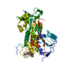
Mass: 18.015 Da / Num. of mol.: 24 / Source method: isolated from a natural source / Formula: H2O

-
Experimental details

-
Experiment

ExperimentMethod: X-RAY DIFFRACTION / Number of used crystals: 1

-
Sample preparation

CrystalDensity Matthews: 3.15 Å3/Da / Density % sol: 60.94 %
Crystal growTemperature: 293.15 K / Method: vapor diffusion, sitting drop / Details: 2.0M Ammonium Sulfate, 0.1M Bicine, pH9.0

-
Data collection

DiffractionMean temperature: 100 K
Diffraction sourceSource: SYNCHROTRON / Site: Diamond / Beamline: I04-1 / Wavelength: 0.987 Å
DetectorType: DECTRIS PILATUS 2M / Detector: PIXEL / Date: Jul 6, 2013
RadiationProtocol: SINGLE WAVELENGTH / Monochromatic (M) / Laue (L): M / Scattering type: x-ray
Radiation wavelengthWavelength: 0.987 Å / Relative weight: 1
ReflectionResolution: 2.3→46.8 Å / Num. obs: 27827 / % possible obs: 99.9 % / Redundancy: 6.5 % / Rmerge(I) obs: 0.057 / Net I/σ(I): 16.6
Reflection shellResolution: 2.3→2.38 Å / Rmerge(I) obs: 0.702

-
Processing

Software
NameVersionClassification
PHENIX(1.10.1_2155: ???)refinement
iMOSFLMdata reduction
Aimlessdata scaling
PHASERphasing
RefinementMethod to determine structure: MOLECULAR REPLACEMENT
Starting model: 2WXW
Resolution: 2.3→46.8 Å / Cross valid method: NONE / σ(F): 1.36 / Phase error: 28.8
RfactorNum. reflection% reflectionSelection details
Rfree0.1889 1402 5.05 %Random selection
Rwork0.1699 ---
obs0.1726 27786 99.89 %-
Solvent computationShrinkage radii: 0.9 Å / VDW probe radii: 1.11 Å
Refinement stepCycle: LAST / Resolution: 2.3→46.8 Å
ProteinNucleic acidLigandSolventTotal
Num. atoms3256 0 39 24 3319
Refine LS restraints
Refine-IDTypeDev idealNumber
X-RAY DIFFRACTIONf_bond_d0.0023371
X-RAY DIFFRACTIONf_angle_d0.4834593
X-RAY DIFFRACTIONf_dihedral_angle_d12.7952004
X-RAY DIFFRACTIONf_chiral_restr0.04549
X-RAY DIFFRACTIONf_plane_restr0.003578
LS refinement shell
Resolution (Å)Rfactor RfreeNum. reflection RfreeRfactor RworkNum. reflection RworkRefine-ID% reflection obs (%)
2.3004-2.38260.28361450.2672614X-RAY DIFFRACTION95
2.3826-2.47790.26541530.24692631X-RAY DIFFRACTION94
2.4779-2.59070.25011350.24032589X-RAY DIFFRACTION95
2.5907-2.72720.23921370.23412684X-RAY DIFFRACTION95
2.7272-2.8980.25981400.22972604X-RAY DIFFRACTION95
2.898-3.12160.21781330.20322649X-RAY DIFFRACTION95
3.1216-3.43550.20651420.18762633X-RAY DIFFRACTION95
3.4355-3.9320.19531380.16412626X-RAY DIFFRACTION95
3.932-4.95150.13361450.12422659X-RAY DIFFRACTION95
4.9515-34.31540.1721300.14742684X-RAY DIFFRACTION95
Refinement TLS params.

Method: refined / Refine-ID: X-RAY DIFFRACTION

IDL112)L122)L132)L222)L232)L332)S11 (Å °)S12 (Å °)S13 (Å °)S21 (Å °)S22 (Å °)S23 (Å °)S31 (Å °)S32 (Å °)S33 (Å °)T112)T122)T132)T222)T232)T332)Origin x (Å)Origin y (Å)Origin z (Å)
12.8102-0.2065-0.31742.76060.4713.9540.08370.2024-0.31880.1725-0.08520.3131-0.1992-0.37310.01280.31560.04110.06390.33790.00650.308413.010161.5337-1.5218
23.5253-0.0859-0.49723.33170.1853.27690.22430.4570.10560.0189-0.11220.349-0.566-0.3687-0.06270.34630.06350.05380.34210.01250.277814.824967.5829-5.9576
32.6450.8215-0.50376.8875-4.67993.18420.26050.11671.0121-0.20120.0256-0.7645-1.22870.6258-0.14921.0658-0.17510.35040.5367-0.0850.715325.786581.84191.8029
43.8966-0.6454-2.6073.82191.37593.9079-0.2069-0.1602-0.68620.04590.1065-0.36420.17850.54860.10950.30780.02080.00780.4740.01010.446137.99554.5145-11.5219
54.5586-0.7569-1.95033.29560.62863.5870.15820.17590.0197-0.0488-0.0086-0.0894-0.48590.2676-0.12060.3493-0.03560.0150.368-0.00140.241129.21264.636-8.7306
Refinement TLS group
IDRefine-IDRefine TLS-IDSelection details
1X-RAY DIFFRACTION1chain 'A' and (resid 1 through 86 )
2X-RAY DIFFRACTION2chain 'A' and (resid 87 through 197 )
3X-RAY DIFFRACTION3chain 'A' and (resid 198 through 242 )
4X-RAY DIFFRACTION4chain 'A' and (resid 243 through 339 )
5X-RAY DIFFRACTION5chain 'A' and (resid 340 through 450 )

+
About Yorodumi

-
News

-
Feb 9, 2022. New format data for meta-information of EMDB entries

New format data for meta-information of EMDB entries

  • Version 3 of the EMDB header file is now the official format.
  • The previous official version 1.9 will be removed from the archive.

Related info.:EMDB header

External links:wwPDB to switch to version 3 of the EMDB data model

-
Aug 12, 2020. Covid-19 info

Covid-19 info

URL: https://pdbj.org/emnavi/covid19.php

New page: Covid-19 featured information page in EM Navigator.

Related info.:Covid-19 info / Mar 5, 2020. Novel coronavirus structure data

+
Mar 5, 2020. Novel coronavirus structure data

Novel coronavirus structure data

Related info.:Yorodumi Speices / Aug 12, 2020. Covid-19 info

External links:COVID-19 featured content - PDBj / Molecule of the Month (242):Coronavirus Proteases

+
Jan 31, 2019. EMDB accession codes are about to change! (news from PDBe EMDB page)

EMDB accession codes are about to change! (news from PDBe EMDB page)

  • The allocation of 4 digits for EMDB accession codes will soon come to an end. Whilst these codes will remain in use, new EMDB accession codes will include an additional digit and will expand incrementally as the available range of codes is exhausted. The current 4-digit format prefixed with “EMD-” (i.e. EMD-XXXX) will advance to a 5-digit format (i.e. EMD-XXXXX), and so on. It is currently estimated that the 4-digit codes will be depleted around Spring 2019, at which point the 5-digit format will come into force.
  • The EM Navigator/Yorodumi systems omit the EMD- prefix.

Related info.:Q: What is EMD? / ID/Accession-code notation in Yorodumi/EM Navigator

External links:EMDB Accession Codes are Changing Soon! / Contact to PDBj

+
Jul 12, 2017. Major update of PDB

Major update of PDB

  • wwPDB released updated PDB data conforming to the new PDBx/mmCIF dictionary.
  • This is a major update changing the version number from 4 to 5, and with Remediation, in which all the entries are updated.
  • In this update, many items about electron microscopy experimental information are reorganized (e.g. em_software).
  • Now, EM Navigator and Yorodumi are based on the updated data.

External links:wwPDB Remediation / Enriched Model Files Conforming to OneDep Data Standards Now Available in the PDB FTP Archive

-
Yorodumi

Thousand views of thousand structures

  • Yorodumi is a browser for structure data from EMDB, PDB, SASBDB, etc.
  • This page is also the successor to EM Navigator detail page, and also detail information page/front-end page for Omokage search.
  • The word "yorodu" (or yorozu) is an old Japanese word meaning "ten thousand". "mi" (miru) is to see.

Related info.:EMDB / PDB / SASBDB / Comparison of 3 databanks / Yorodumi Search / Aug 31, 2016. New EM Navigator & Yorodumi / Yorodumi Papers / Jmol/JSmol / Function and homology information / Changes in new EM Navigator and Yorodumi

Read more