 Movie
Movie Controller

[English] 日本語
 Yorodumi
- PDB-7laq: Crystal structure of Campylobacter jejuni Cj0843c lytic transglyc... -
+ Open data

- Basic information
| Entry | Database: PDB / ID: 7laq | ||||||
|---|---|---|---|---|---|---|---|
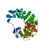
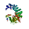
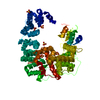
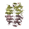
| Title | Crystal structure of Campylobacter jejuni Cj0843c lytic transglycosylase in complex with N,N'-diacetylchitobiose | ||||||
 Components Components | Lytic transglycosylase domain-containing protein | ||||||
 Keywords Keywords | LYASE/LYASE INHIBITOR / inhibitor / complex / substrate analog / LYASE / LYASE-LYASE INHIBITOR complex | ||||||
| Function / homology |  Function and homology information Function and homology informationpeptidoglycan lytic transglycosylase activity / peptidoglycan metabolic process / membrane Similarity search - Function | ||||||
| Biological species |   Campylobacter jejuni (Campylobacter) Campylobacter jejuni (Campylobacter) | ||||||
| Method |  X-RAY DIFFRACTION / SYNCHROTRON / MOLECULAR REPLACEMENT / Resolution: 2.58 Å X-RAY DIFFRACTION / SYNCHROTRON / MOLECULAR REPLACEMENT / Resolution: 2.58 Å | ||||||
 Authors Authors | van den Akker, F. / Kumar, V. | ||||||
| Funding support |  United States, 1items United States, 1items
| ||||||
 Citation Citation | Journal: Biochemistry / Year: 2021 Title: Turnover Chemistry and Structural Characterization of the Cj0843c Lytic Transglycosylase of Campylobacter jejuni . Authors: Kumar, V. / Mathure, S.A. / Lee, M. / Boorman, J. / Zeng, X. / Lin, J. / Hesek, D. / Lastochkin, E. / Mobashery, S. / van den Akker, F. | ||||||
| History |
|
- Structure visualization
| Structure viewer | Molecule: MolmilJmol/JSmol |
|---|
- Downloads & links
-Download
| PDBx/mmCIF format | 7laq.cif.gz | 128.2 KB | Display | PDBx/mmCIF format |
|---|---|---|---|---|
| PDB format | pdb7laq.ent.gz | 97.5 KB | Display | PDB format |
| PDBx/mmJSON format | 7laq.json.gz | Tree view | PDBx/mmJSON format | |
| Others |  Other downloads Other downloads |
-Validation report
| Arichive directory | https://data.pdbj.org/pub/pdb/validation_reports/la/7laqftp://data.pdbj.org/pub/pdb/validation_reports/la/7laq | HTTPS FTP |
|---|
-Related structure data
| Related structure data |  7lamC  6cf9S C: citing same article ( S: Starting model for refinement |
|---|---|
| Similar structure data |


-Links
 PDBj
PDBj- Assembly
| Deposited unit | 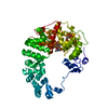
| ||||||||
|---|---|---|---|---|---|---|---|---|---|
| 1 |
| ||||||||
| Unit cell |
| ||||||||
| Components on special symmetry positions |
|
-Components
| #1: Protein | Mass: 63644.652 Da / Num. of mol.: 1 Source method: isolated from a genetically manipulated source Source: (gene. exp.) Campylobacter jejuni (Campylobacter)Gene: A0X18_03655, A0X31_01845, A9372_04065, APU74_07480, B1933_07625, BBS05_05650, BCB47_05730, BED64_07690, C3I22_05260, C3I27_06990, C3I35_05175, C7N26_08315, CJ274_08505, CV323_05370, CWD74_ ...Gene: A0X18_03655, A0X31_01845, A9372_04065, APU74_07480, B1933_07625, BBS05_05650, BCB47_05730, BED64_07690, C3I22_05260, C3I27_06990, C3I35_05175, C7N26_08315, CJ274_08505, CV323_05370, CWD74_05755, D0W34_08355, D4Q41_06000, D5I02_03510, D6H33_08420, DPG08_07275, DUX97_06720, DUY05_08000, DWS06_05135, DYE84_06365, E7R20_02805, EAX31_05720, EC071_05580, F0166_06250, F6982_06065, F7521_08930, F7J47_05300, F7U05_05765, F8803_06810, F9778_04965, FCR67_06545, FPD29_09025, FPT92_05795, FV831_05555, FV933_07400, FVI24_07230, FVM64_02425, FVM77_01160, FW220_07595, FW611_08035, FW976_03985, FWA25_07655, FY101_03890, FZ445_05005, FZ878_07970, FZW01_06295, G3M94_001083, GAX22_07420, GCI37_01695, GI172_07425, GIT96_06475, GJ442_07755, GK482_04850, GK486_00445, GL007_04270, GL031_07375, GM780_07860, GMG42_07100, GN862_07225, GNO13_05645, GNO32_03000, GPD80_07165, GRH33_07480, GRM82_06860, GRO30_05060, GRS20_08245, GSH24_05325, GTJ31_06080, GTV40_06285, GTV58_05620, GWW45_06140, GY415_000965, GZ489_001419, GZ499_001624, GZ502_000976, GZ518_001219 Production host: | ||||||||||
|---|---|---|---|---|---|---|---|---|---|---|---|
| #2: Polysaccharide | | #3: Chemical | ChemComp-CIT / |   Mass: 192.124 Da / Num. of mol.: 1 / Source method: obtained synthetically / Formula: C6H8O7 Mass: 192.124 Da / Num. of mol.: 1 / Source method: obtained synthetically / Formula: C6H8O7#4: Chemical | ChemComp-ACT / |   Mass: 59.044 Da / Num. of mol.: 1 / Source method: obtained synthetically / Formula: C2H3O2 Mass: 59.044 Da / Num. of mol.: 1 / Source method: obtained synthetically / Formula: C2H3O2#5: Water | ChemComp-HOH / |  Mass: 18.015 Da / Num. of mol.: 193 / Source method: isolated from a natural source / Formula: H2O Mass: 18.015 Da / Num. of mol.: 193 / Source method: isolated from a natural source / Formula: H2OHas ligand of interest | Y | Has protein modification | Y | |
-Experimental details
-Experiment
| Experiment | Method: X-RAY DIFFRACTION / Number of used crystals: 1 |
|---|
- Sample preparation
| Crystal | Density Matthews: 3.63 Å3/Da / Density % sol: 66.11 % |
|---|---|
| Crystal grow | Temperature: 293 K / Method: vapor diffusion, sitting drop Details: 100 sodium citrate buffer pH 5.1-6.0, and 25-33% PEG 600. The protein was concentrated to 10 mg/ml in 10 mM HEPES pH 8.0 and 200 mM ammonium sulfate PH range: 5.1-6.0 |
-Data collection
| Diffraction | Mean temperature: 100 K / Serial crystal experiment: N |
|---|---|
| Diffraction source | Source: SYNCHROTRON / Site: NSLS / Beamline: X17B1 / Wavelength: 0.97935 Å |
| Detector | Type: DECTRIS EIGER X 16M / Detector: PIXEL / Date: Aug 29, 2020 |
| Radiation | Protocol: SINGLE WAVELENGTH / Monochromatic (M) / Laue (L): M / Scattering type: x-ray |
| Radiation wavelength | Wavelength: 0.97935 Å / Relative weight: 1 |
| Reflection | Resolution: 2.58→29.52 Å / Num. obs: 28913 / % possible obs: 99.5 % / Redundancy: 13.9 % / CC1/2: 0.997 / Rmerge(I) obs: 0.17 / Net I/σ(I): 13 |
| Reflection shell | Resolution: 2.58→2.65 Å / Rmerge(I) obs: 1.018 / Num. unique obs: 2029 / CC1/2: 0.782 |
- Processing
| Software |
| ||||||||||||||||||||||||||||||||||||||||||||||||||||||||||||
|---|---|---|---|---|---|---|---|---|---|---|---|---|---|---|---|---|---|---|---|---|---|---|---|---|---|---|---|---|---|---|---|---|---|---|---|---|---|---|---|---|---|---|---|---|---|---|---|---|---|---|---|---|---|---|---|---|---|---|---|---|---|
| Refinement | Method to determine structure: MOLECULAR REPLACEMENT Starting model: 6CF9 Resolution: 2.58→29.5 Å / Cor.coef. Fo:Fc: 0.955 / Cor.coef. Fo:Fc free: 0.929 / SU B: 8.497 / SU ML: 0.178 / Cross valid method: THROUGHOUT / σ(F): 0 / ESU R: 0.326 / ESU R Free: 0.246 / Stereochemistry target values: MAXIMUM LIKELIHOOD Details: HYDROGENS HAVE BEEN ADDED IN THE RIDING POSITIONS U VALUES : REFINED INDIVIDUALLY
| ||||||||||||||||||||||||||||||||||||||||||||||||||||||||||||
| Solvent computation | Ion probe radii: 0.8 Å / Shrinkage radii: 0.8 Å / VDW probe radii: 1.2 Å / Solvent model: MASK | ||||||||||||||||||||||||||||||||||||||||||||||||||||||||||||
| Displacement parameters | Biso max: 147.66 Å2 / Biso mean: 42.196 Å2 / Biso min: 5.97 Å2
| ||||||||||||||||||||||||||||||||||||||||||||||||||||||||||||
| Refinement step | Cycle: final / Resolution: 2.58→29.5 Å
| ||||||||||||||||||||||||||||||||||||||||||||||||||||||||||||
| Refine LS restraints |
| ||||||||||||||||||||||||||||||||||||||||||||||||||||||||||||
| LS refinement shell | Resolution: 2.583→2.65 Å / Rfactor Rfree error: 0 / Total num. of bins used: 20
|
