 Movie
Movie Controller

[English] 日本語
 Yorodumi
- PDB-6cf8: Crystal structure of Cj0843 lytic transglycosylase of Campylobact... -
+ Open data

- Basic information
| Entry | Database: PDB / ID: 6cf8 | ||||||
|---|---|---|---|---|---|---|---|
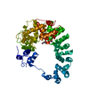
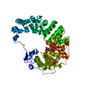
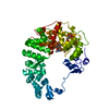
| Title | Crystal structure of Cj0843 lytic transglycosylase of Campylobacter jejuni at 1.87A resolution | ||||||
 Components Components | Lytic transglycosylase | ||||||
 Keywords Keywords | HYDROLASE | ||||||
| Function / homology | :  Function and homology information Function and homology information | ||||||
| Biological species |   Campylobacter jejuni (Campylobacter) Campylobacter jejuni (Campylobacter) | ||||||
| Method |  X-RAY DIFFRACTION / SYNCHROTRON / SAD / Resolution: 1.87 Å X-RAY DIFFRACTION / SYNCHROTRON / SAD / Resolution: 1.87 Å | ||||||
 Authors Authors | van den Akker, F. / Kumar, V. / Vijayaraghavan, J. | ||||||
 Citation Citation | Journal: PLoS ONE / Year: 2018 Title: Structural studies and molecular dynamics simulations suggest a processive mechanism of exolytic lytic transglycosylase from Campylobacter jejuni. Authors: Vijayaraghavan, J. / Kumar, V. / Krishnan, N.P. / Kaufhold, R.T. / Zeng, X. / Lin, J. / van den Akker, F. | ||||||
| History |
|
- Structure visualization
| Structure viewer | Molecule: MolmilJmol/JSmol |
|---|
- Downloads & links
-Download
| PDBx/mmCIF format | 6cf8.cif.gz | 129.4 KB | Display | PDBx/mmCIF format |
|---|---|---|---|---|
| PDB format | pdb6cf8.ent.gz | 98.2 KB | Display | PDB format |
| PDBx/mmJSON format | 6cf8.json.gz | Tree view | PDBx/mmJSON format | |
| Others |  Other downloads Other downloads |
-Validation report
| Arichive directory | https://data.pdbj.org/pub/pdb/validation_reports/cf/6cf8ftp://data.pdbj.org/pub/pdb/validation_reports/cf/6cf8 | HTTPS FTP |
|---|
-Related structure data


-Links
 PDBj
PDBj- Assembly
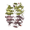
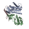
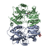
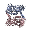
| Deposited unit | 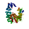
| ||||||||
|---|---|---|---|---|---|---|---|---|---|
| 1 |
| ||||||||
| Unit cell |
| ||||||||
| Components on special symmetry positions |
|
-Components
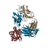
| #1: Protein | Mass: 61487.293 Da / Num. of mol.: 1 / Fragment: residues 18-540 Source method: isolated from a genetically manipulated source Source: (gene. exp.) Campylobacter jejuni (Campylobacter) / Gene: BD28_04025 / Production host: | ||||
|---|---|---|---|---|---|
| #2: Chemical | ChemComp-SO4 /   Mass: 96.063 Da / Num. of mol.: 7 / Source method: obtained synthetically / Formula: SO4 Mass: 96.063 Da / Num. of mol.: 7 / Source method: obtained synthetically / Formula: SO4#3: Water | ChemComp-HOH / |  Mass: 18.015 Da / Num. of mol.: 409 / Source method: isolated from a natural source / Formula: H2O Mass: 18.015 Da / Num. of mol.: 409 / Source method: isolated from a natural source / Formula: H2OHas protein modification | Y | |
-Experimental details
-Experiment
| Experiment | Method: X-RAY DIFFRACTION / Number of used crystals: 1 |
|---|
- Sample preparation
| Crystal | Density Matthews: 2.59 Å3/Da / Density % sol: 52.42 % |
|---|---|
| Crystal grow | Temperature: 298 K / Method: vapor diffusion, sitting drop / pH: 8 Details: 0.2 M lithium sulfate, 0.1 M Tris pH 8.0, and 39% PEG 400 |
-Data collection
| Diffraction | Mean temperature: 100 K |
|---|---|
| Diffraction source | Source: SYNCHROTRON / Site: SSRL  / Beamline: BL9-2 / Wavelength: 0.97946 Å / Beamline: BL9-2 / Wavelength: 0.97946 Å |
| Detector | Type: DECTRIS PILATUS3 S 6M / Detector: PIXEL / Date: Jun 29, 2017 |
| Radiation | Protocol: SINGLE WAVELENGTH / Monochromatic (M) / Laue (L): M / Scattering type: x-ray |
| Radiation wavelength | Wavelength: 0.97946 Å / Relative weight: 1 |
| Reflection | Resolution: 1.87→64.7 Å / Num. obs: 51659 / % possible obs: 98.3 % / Redundancy: 19 % / Net I/σ(I): 16.3 |
| Reflection shell | Resolution: 1.87→1.92 Å |
- Processing
| Software |
| ||||||||||||||||||||||||||||||||||||||||||||||||||||||||||||
|---|---|---|---|---|---|---|---|---|---|---|---|---|---|---|---|---|---|---|---|---|---|---|---|---|---|---|---|---|---|---|---|---|---|---|---|---|---|---|---|---|---|---|---|---|---|---|---|---|---|---|---|---|---|---|---|---|---|---|---|---|---|
| Refinement | Method to determine structure: SAD / Resolution: 1.87→64.7 Å / Cor.coef. Fo:Fc: 0.963 / Cor.coef. Fo:Fc free: 0.941 / SU B: 3.574 / SU ML: 0.104 / Cross valid method: THROUGHOUT / σ(F): 0 / ESU R: 0.144 / ESU R Free: 0.143 Details: HYDROGENS HAVE BEEN ADDED IN THE RIDING POSITIONS U VALUES : REFINED INDIVIDUALLY
| ||||||||||||||||||||||||||||||||||||||||||||||||||||||||||||
| Solvent computation | Ion probe radii: 0.8 Å / Shrinkage radii: 0.8 Å / VDW probe radii: 1.2 Å | ||||||||||||||||||||||||||||||||||||||||||||||||||||||||||||
| Displacement parameters | Biso max: 106.08 Å2 / Biso mean: 42.006 Å2 / Biso min: 22.12 Å2
| ||||||||||||||||||||||||||||||||||||||||||||||||||||||||||||
| Refinement step | Cycle: final / Resolution: 1.87→64.7 Å
| ||||||||||||||||||||||||||||||||||||||||||||||||||||||||||||
| Refine LS restraints |
| ||||||||||||||||||||||||||||||||||||||||||||||||||||||||||||
| LS refinement shell | Resolution: 1.874→1.922 Å / Rfactor Rfree error: 0 / Total num. of bins used: 20
|
