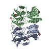
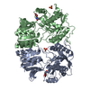
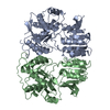
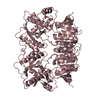
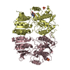
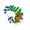
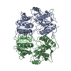
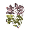
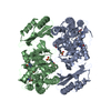
Entry Database : PDB / ID : 3o2jTitle Structure of the GluA2 NTD-dimer interface mutant, N54A Glutamate receptor 2 Keywords / / / / / Function / homology Function Domain/homology Component
/ / / / / / / / / / / / / / / / / / / / / / / / / / / / / / / / / / / / / / / / / / / / / / / / / / / / / / / / / / / / / / / / / / / / / / / / / / / / / / / / / / / / / / / / / / / / / / / / / / / / / / / / / / / / / / / / / / / Biological species Rattus norvegicus (Norway rat)Method / / / Resolution : 1.95 Å Authors Rossmann, M. / Sukumaran, M. / Penn, A.C. / Veprintsev, D.B. / Greger, I.H. Journal : Embo J. / Year : 2011Title : Subunit-selective N-terminal domain associations organize the formation of AMPA receptor heteromersAuthors : Rossmann, M. / Sukumaran, M. / Penn, A.C. / Veprintsev, D.B. / Babu, M.M. / Greger, I.H. History Deposition Jul 22, 2010 Deposition site / Processing site Revision 1.0 Mar 9, 2011 Provider / Type Revision 1.1 Jul 13, 2011 Group Revision 1.2 Jul 29, 2020 Group Data collection / Database references ... Data collection / Database references / Derived calculations / Structure summary Category chem_comp / entity ... chem_comp / entity / pdbx_chem_comp_identifier / pdbx_entity_nonpoly / struct_conn / struct_ref_seq_dif / struct_site / struct_site_gen Item _chem_comp.name / _chem_comp.type ... _chem_comp.name / _chem_comp.type / _entity.pdbx_description / _pdbx_entity_nonpoly.name / _struct_conn.pdbx_leaving_atom_flag / _struct_conn.pdbx_role / _struct_ref_seq_dif.details Description / Provider / Type Revision 1.3 Nov 1, 2023 Group Data collection / Database references ... Data collection / Database references / Refinement description / Structure summary Category chem_comp / chem_comp_atom ... chem_comp / chem_comp_atom / chem_comp_bond / database_2 / pdbx_initial_refinement_model Item / _database_2.pdbx_DOI / _database_2.pdbx_database_accessionRevision 1.4 Nov 6, 2024 Group / Category / pdbx_modification_feature
Show all Show less
 Movie
Movie Controller


 Open data
 Basic information
 Components
 Keywords
 Function and homology information
 X-RAY DIFFRACTION /
  Authors
 Citation
 Structure visualization
 Downloads & links
 Other downloads
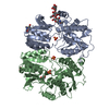


 PDBj
PDBj


 Assembly
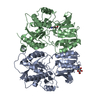
 Homo sapiens (human) / References: UniProt: P19491
 Type: D-saccharide, beta linking / Mass: 221.208 Da / Num. of mol.: 2
 Mass: 18.015 Da / Num. of mol.: 405 / Source method: isolated from a natural source / Formula: H2O
 Sample preparation
 / Beamline: I03 / Wavelength: 0.9763 Å
 Processing