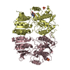
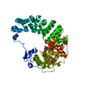
Entry Database : PDB / ID : 3h5wTitle Crystal structure of the GluR2-ATD in space group P212121 without solvent Glutamate receptor 2 Keywords / / / / / / / / / / / / / / / / / / / Function / homology Function Domain/homology Component
/ / / / / / / / / / / / / / / / / / / / / / / / / / / / / / / / / / / / / / / / / / / / / / / / / / / / / / / / / / / / / / / / / / / / / / / / / / / / / / / / / / / / / / / / / / / / / / / / / / / / / / / / / / / / / / / / / / / Biological species Rattus norvegicus (Norway rat)Method / / / Resolution : 2.686 Å Authors Jin, R. / Singh, S.K. / Gu, S. / Furukawa, H. / Sobolevsky, A. / Zhou, J. / Jin, Y. / Gouaux, E. Journal : Embo J. / Year : 2009Title : Crystal structure and association behaviour of the GluR2 amino-terminal domain.Authors : Jin, R. / Singh, S.K. / Gu, S. / Furukawa, H. / Sobolevsky, A.I. / Zhou, J. / Jin, Y. / Gouaux, E. History Deposition Apr 22, 2009 Deposition site / Processing site Revision 1.0 Jun 9, 2009 Provider / Type Revision 1.1 Jul 13, 2011 Group Revision 1.2 Sep 6, 2023 Group / Database references / Refinement descriptionCategory chem_comp_atom / chem_comp_bond ... chem_comp_atom / chem_comp_bond / database_2 / pdbx_initial_refinement_model / struct_ref_seq_dif Item / _database_2.pdbx_database_accession / _struct_ref_seq_dif.detailsRevision 1.3 Oct 9, 2024 Group / Category / pdbx_modification_feature
Show all Show less
 Movie
Movie Controller

 Yorodumi
 Open data
 Basic information
 Components
 Keywords
 Function and homology information
 X-RAY DIFFRACTION /
  Authors
 Citation
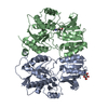
 Structure visualization
 Downloads & links
 Other downloads
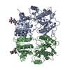

 PDBj
PDBj


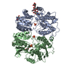
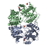
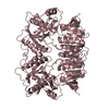
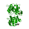
 Assembly
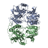

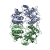
 Spodoptera frugiperda (fall armyworm) / References: UniProt: P19491
 Sample preparation
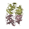
 / Beamline: 5.0.2 / Wavelength: 1 Å
 Processing