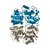
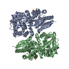
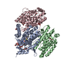
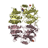
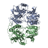
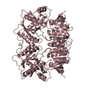
[English] 日本語
Yorodumi
- PDB-3h5v: Crystal structure of the GluR2-ATD -

+
Open data


ID or keywords:

Loading...

-
Basic information

Entry
Database: PDB / ID: 3h5v
TitleCrystal structure of the GluR2-ATD
ComponentsGlutamate receptor 2
KeywordsTRANSPORT PROTEIN / Glutamate receptor / Ligand-gated ion channel / synapse / Cell junction / Cell membrane / Endoplasmic reticulum / Glycoprotein / Ion transport / Ionic channel / Lipoprotein / Membrane / Palmitate / Phosphoprotein / Postsynaptic cell membrane / Receptor / RNA editing / Transmembrane / Transport
Function / homology
Function and homology information


spine synapse / dendritic spine neck / dendritic spine head / cellular response to amine stimulus / perisynaptic space / Activation of AMPA receptors / ligand-gated monoatomic cation channel activity / AMPA glutamate receptor activity / response to lithium ion / Trafficking of GluR2-containing AMPA receptors ...spine synapse / dendritic spine neck / dendritic spine head / cellular response to amine stimulus / perisynaptic space / Activation of AMPA receptors / ligand-gated monoatomic cation channel activity / AMPA glutamate receptor activity / response to lithium ion / Trafficking of GluR2-containing AMPA receptors / kainate selective glutamate receptor activity / cellular response to glycine / AMPA glutamate receptor complex / extracellularly glutamate-gated ion channel activity / immunoglobulin binding / asymmetric synapse / ionotropic glutamate receptor complex / conditioned place preference / regulation of receptor recycling / glutamate receptor binding / Unblocking of NMDA receptors, glutamate binding and activation / positive regulation of synaptic transmission / regulation of synaptic transmission, glutamatergic / response to fungicide / cytoskeletal protein binding / glutamate-gated receptor activity / regulation of long-term synaptic depression / cellular response to brain-derived neurotrophic factor stimulus / extracellular ligand-gated monoatomic ion channel activity / glutamate-gated calcium ion channel activity / presynaptic active zone membrane / somatodendritic compartment / dendrite membrane / ionotropic glutamate receptor binding / ligand-gated monoatomic ion channel activity involved in regulation of presynaptic membrane potential / ionotropic glutamate receptor signaling pathway / dendrite cytoplasm / synaptic membrane / dendritic shaft / SNARE binding / transmitter-gated monoatomic ion channel activity involved in regulation of postsynaptic membrane potential / synaptic transmission, glutamatergic / PDZ domain binding / protein tetramerization / establishment of protein localization / postsynaptic density membrane / cerebral cortex development / modulation of chemical synaptic transmission / receptor internalization / Schaffer collateral - CA1 synapse / terminal bouton / synaptic vesicle / synaptic vesicle membrane / signaling receptor activity / presynapse / amyloid-beta binding / growth cone / presynaptic membrane / scaffold protein binding / perikaryon / dendritic spine / chemical synaptic transmission / postsynaptic membrane / neuron projection / postsynaptic density / axon / external side of plasma membrane / neuronal cell body / synapse / dendrite / protein kinase binding / protein-containing complex binding / glutamatergic synapse / cell surface / endoplasmic reticulum / protein-containing complex / identical protein binding / membrane / plasma membrane
Similarity search - Function
Response regulator / Ionotropic glutamate receptor, metazoa / Ligated ion channel L-glutamate- and glycine-binding site / Ionotropic glutamate receptor, L-glutamate and glycine-binding domain / Ligated ion channel L-glutamate- and glycine-binding site / Ligand-gated ion channel / : / Ionotropic glutamate receptor / Eukaryotic homologues of bacterial periplasmic substrate binding proteins. / Receptor, ligand binding region ...Response regulator / Ionotropic glutamate receptor, metazoa / Ligated ion channel L-glutamate- and glycine-binding site / Ionotropic glutamate receptor, L-glutamate and glycine-binding domain / Ligated ion channel L-glutamate- and glycine-binding site / Ligand-gated ion channel / : / Ionotropic glutamate receptor / Eukaryotic homologues of bacterial periplasmic substrate binding proteins. / Receptor, ligand binding region / Receptor family ligand binding region / Periplasmic binding protein-like I / Rossmann fold / 3-Layer(aba) Sandwich / Alpha Beta
Similarity search - Domain/homology
Glutamate receptor 2
Similarity search - Component
Biological speciesRattus norvegicus (Norway rat)
MethodX-RAY DIFFRACTION / SYNCHROTRON / SAD / Resolution: 2.33 Å
AuthorsJin, R. / Singh, S.K. / Gu, S. / Furukawa, H. / Sobolevsky, A. / Zhou, J. / Jin, Y. / Gouaux, E.
CitationJournal: Embo J. / Year: 2009
Title: Crystal structure and association behaviour of the GluR2 amino-terminal domain.
Authors: Jin, R. / Singh, S.K. / Gu, S. / Furukawa, H. / Sobolevsky, A.I. / Zhou, J. / Jin, Y. / Gouaux, E.
History
DepositionApr 22, 2009Deposition site: RCSB / Processing site: RCSB
Revision 1.0Jun 9, 2009Provider: repository / Type: Initial release
Revision 1.1Jul 13, 2011Group: Non-polymer description / Version format compliance
Revision 2.0Jul 29, 2020Group: Advisory / Atomic model ...Advisory / Atomic model / Data collection / Database references / Derived calculations / Structure summary
Category: atom_site / chem_comp ...atom_site / chem_comp / entity / pdbx_branch_scheme / pdbx_chem_comp_identifier / pdbx_entity_branch / pdbx_entity_branch_descriptor / pdbx_entity_branch_link / pdbx_entity_branch_list / pdbx_entity_nonpoly / pdbx_nonpoly_scheme / pdbx_struct_assembly_gen / pdbx_struct_special_symmetry / pdbx_unobs_or_zero_occ_atoms / struct_asym / struct_conn / struct_ref_seq_dif / struct_site / struct_site_gen
Item: _atom_site.auth_asym_id / _atom_site.auth_seq_id ..._atom_site.auth_asym_id / _atom_site.auth_seq_id / _atom_site.label_asym_id / _chem_comp.name / _chem_comp.type / _entity.formula_weight / _entity.pdbx_description / _entity.pdbx_number_of_molecules / _entity.type / _pdbx_struct_assembly_gen.asym_id_list / _pdbx_struct_special_symmetry.label_asym_id / _struct_conn.pdbx_leaving_atom_flag / _struct_conn.pdbx_role / _struct_conn.ptnr1_auth_asym_id / _struct_conn.ptnr1_auth_seq_id / _struct_conn.ptnr1_label_asym_id / _struct_conn.ptnr2_auth_asym_id / _struct_conn.ptnr2_auth_seq_id / _struct_conn.ptnr2_label_asym_id / _struct_ref_seq_dif.details
Description: Carbohydrate remediation / Provider: repository / Type: Remediation
Revision 2.1Nov 6, 2024Group: Advisory / Data collection ...Advisory / Data collection / Database references / Structure summary
Category: chem_comp / chem_comp_atom ...chem_comp / chem_comp_atom / chem_comp_bond / database_2 / pdbx_entry_details / pdbx_modification_feature / pdbx_unobs_or_zero_occ_atoms
Item: _chem_comp.pdbx_synonyms / _database_2.pdbx_DOI / _database_2.pdbx_database_accession

-
Structure visualization

Structure viewerMolecule:
MolmilJmol/JSmol

Downloads & links

-
Assembly

Deposited unit
A: Glutamate receptor 2
B: Glutamate receptor 2
C: Glutamate receptor 2
hetero molecules


Theoretical massNumber of molelcules
Total (without water)134,3775
Polymers133,5283
Non-polymers8492
Water6,630368
1
A: Glutamate receptor 2
B: Glutamate receptor 2
hetero molecules


Theoretical massNumber of molelcules
Total (without water)89,8674
Polymers89,0192
Non-polymers8492
Water362
TypeNameSymmetry operationNumber
identity operation1_555x,y,z1
Buried area3840 Å2
ΔGint3 kcal/mol
Surface area32510 Å2
MethodPISA
2
C: Glutamate receptor 2

C: Glutamate receptor 2


Theoretical massNumber of molelcules
Total (without water)89,0192
Polymers89,0192
Non-polymers00
Water362
TypeNameSymmetry operationNumber
identity operation1_555x,y,z1
crystal symmetry operation2_565-x,-y+1,z1
Buried area2730 Å2
ΔGint-16 kcal/mol
Surface area31860 Å2
MethodPISA
Unit cell
Length a, b, c (Å)64.003, 362.993, 61.434
Angle α, β, γ (deg.)90.00, 90.00, 90.00
Int Tables number18
Space group name H-MP21212
Components on special symmetry positions
IDModelComponents
11C-392-

HOH

-
Components

#1: Protein Glutamate receptor 2 / GluR-2 / GluR-B / GluR-K2 / Glutamate receptor ionotropic / AMPA 2 / AMPA-selective glutamate receptor 2


Mass: 44509.250 Da / Num. of mol.: 3 / Fragment: UNP residues 21-404
Source method: isolated from a genetically manipulated source
Source: (gene. exp.) Rattus norvegicus (Norway rat) / Gene: Gria2, Glur2 / Production host: Spodoptera frugiperda (fall armyworm) / References: UniProt: P19491
#2: Polysaccharide 2-acetamido-2-deoxy-beta-D-glucopyranose-(1-4)-2-acetamido-2-deoxy-beta-D-glucopyranose


Type: oligosaccharide / Mass: 424.401 Da / Num. of mol.: 2
Source method: isolated from a genetically manipulated source
DescriptorTypeProgram
DGlcpNAcb1-4DGlcpNAcb1-Glycam Condensed SequenceGMML 1.0
WURCS=2.0/1,2,1/[a2122h-1b_1-5_2*NCC/3=O]/1-1/a4-b1WURCSPDB2Glycan 1.1.0
[]{[(4+1)][b-D-GlcpNAc]{[(4+1)][b-D-GlcpNAc]{}}}LINUCSPDB-CARE
#3: Water ChemComp-HOH / water


Mass: 18.015 Da / Num. of mol.: 368 / Source method: isolated from a natural source / Formula: H2O
Has protein modificationY

-
Experimental details

-
Experiment

ExperimentMethod: X-RAY DIFFRACTION / Number of used crystals: 1

-
Sample preparation

CrystalDensity Matthews: 2.67 Å3/Da / Density % sol: 53.97 %
Crystal growTemperature: 277 K / Method: vapor diffusion, hanging drop / pH: 8
Details: 14-16% PEG3350, 100mM TrisHCl, 10mM MgCl2, pH 8.0, VAPOR DIFFUSION, HANGING DROP, temperature 277K

-
Data collection

DiffractionMean temperature: 100 K
Diffraction sourceSource: SYNCHROTRON / Site: APS / Beamline: 24-ID-C / Wavelength: 1 Å
DetectorType: ADSC QUANTUM 315 / Detector: CCD / Date: Jan 30, 2008
RadiationMonochromator: Si(111) / Protocol: SINGLE WAVELENGTH / Monochromatic (M) / Laue (L): M / Scattering type: x-ray
Radiation wavelengthWavelength: 1 Å / Relative weight: 1
ReflectionResolution: 2.33→50 Å / Num. obs: 61441 / % possible obs: 99.9 % / Observed criterion σ(F): 0 / Observed criterion σ(I): 0 / Redundancy: 7.3 % / Rmerge(I) obs: 0.061 / Net I/σ(I): 35.8
Reflection shellResolution: 2.33→2.41 Å / Redundancy: 7.4 % / Rmerge(I) obs: 0.605 / Mean I/σ(I) obs: 2.2 / % possible all: 99.9

-
Processing

Software
NameVersionClassification
HKL-2000data collection
SOLVEphasing
PHENIX(phenix.refine)refinement
HKL-2000data reduction
HKL-2000data scaling
RefinementMethod to determine structure: SAD / Resolution: 2.33→19.912 Å / SU ML: 0.34 / σ(F): 1.33 / Phase error: 23.2 / Stereochemistry target values: ML
RfactorNum. reflection% reflection
Rfree0.2355 5084 8.14 %
Rwork0.1859 --
obs0.1899 61441 99.94 %
Solvent computationShrinkage radii: 0.9 Å / VDW probe radii: 1.11 Å / Solvent model: FLAT BULK SOLVENT MODEL / Bsol: 72.811 Å2 / ksol: 0.316 e/Å3
Refinement stepCycle: LAST / Resolution: 2.33→19.912 Å
ProteinNucleic acidLigandSolventTotal
Num. atoms8968 0 56 368 9392
Refine LS restraints
Refine-IDTypeDev idealNumber
X-RAY DIFFRACTIONf_bond_d0.0089213
X-RAY DIFFRACTIONf_angle_d1.08912459
X-RAY DIFFRACTIONf_dihedral_angle_d17.8723338
X-RAY DIFFRACTIONf_chiral_restr0.0741380
X-RAY DIFFRACTIONf_plane_restr0.0041603
LS refinement shell
Resolution (Å)Rfactor RfreeNum. reflection RfreeRfactor RworkNum. reflection RworkRefine-ID% reflection obs (%)
2.33-2.35640.30661580.23611912X-RAY DIFFRACTION100
2.3564-2.38410.26631780.21291853X-RAY DIFFRACTION100
2.3841-2.41310.27821750.19231859X-RAY DIFFRACTION100
2.4131-2.44360.27181660.19591895X-RAY DIFFRACTION100
2.4436-2.47570.28321670.20091889X-RAY DIFFRACTION100
2.4757-2.50960.29071570.19211848X-RAY DIFFRACTION100
2.5096-2.54540.28871680.19341920X-RAY DIFFRACTION100
2.5454-2.58330.26971470.18291899X-RAY DIFFRACTION100
2.5833-2.62360.2431760.1811864X-RAY DIFFRACTION100
2.6236-2.66650.25481510.181953X-RAY DIFFRACTION100
2.6665-2.71230.24741760.18291835X-RAY DIFFRACTION100
2.7123-2.76150.23411660.17551903X-RAY DIFFRACTION100
2.7615-2.81450.25971690.19061889X-RAY DIFFRACTION100
2.8145-2.87180.26451730.18731876X-RAY DIFFRACTION100
2.8718-2.93410.25021620.18961922X-RAY DIFFRACTION100
2.9341-3.00210.25031700.18591872X-RAY DIFFRACTION100
3.0021-3.07690.25921680.18371953X-RAY DIFFRACTION100
3.0769-3.15980.29321910.19981843X-RAY DIFFRACTION100
3.1598-3.25240.25541740.21031922X-RAY DIFFRACTION100
3.2524-3.35690.21281610.19321898X-RAY DIFFRACTION100
3.3569-3.47620.25761720.19341907X-RAY DIFFRACTION100
3.4762-3.61460.24421750.18671926X-RAY DIFFRACTION100
3.6146-3.77810.23271610.18121923X-RAY DIFFRACTION100
3.7781-3.97580.24421800.17631910X-RAY DIFFRACTION100
3.9758-4.22270.21681720.15891940X-RAY DIFFRACTION100
4.2227-4.54510.16751620.1481938X-RAY DIFFRACTION100
4.5451-4.9960.18761680.14971965X-RAY DIFFRACTION100
4.996-5.7040.2231810.1781959X-RAY DIFFRACTION100
5.704-7.13110.22711780.19612008X-RAY DIFFRACTION100
7.1311-19.91320.21691820.18352085X-RAY DIFFRACTION99
Refinement TLS params.

Method: refined / Refine-ID: X-RAY DIFFRACTION

IDL112)L122)L132)L222)L232)L332)S11 (Å °)S12 (Å °)S13 (Å °)S21 (Å °)S22 (Å °)S23 (Å °)S31 (Å °)S32 (Å °)S33 (Å °)T112)T122)T132)T222)T232)T332)Origin x (Å)Origin y (Å)Origin z (Å)
10.7921-0.1356-0.72451.9607-0.36932.6411-0.05320.1327-0.05610.0677-0.0824-0.0925-0.1679-0.0940.10780.14820.0073-0.05910.27880.05390.23358.9746105.875130.5343
22.0305-0.0097-0.46780.7837-1.08433.52720.0717-0.13240.1250.35-0.5804-0.2202-0.77920.52930.37690.8137-0.0818-0.13940.44170.06910.348217.4609121.995550.3735
30.8817-0.0545-0.51782.0515-0.30372.136-0.12680.0703-0.07520.2169-0.010.3169-0.0615-0.48130.08750.15230.03950.02720.39850.01540.30651.0773102.711537.6585
41.0568-0.3979-0.46582.6815-0.14332.18450.01960.06340.0981-0.4979-0.1967-0.2453-0.20090.21970.0850.335-0.0420.06060.34270.14660.269230.225118.927613.1791
51.27840.2478-0.57790.2144-2.73612.5933-0.0142-0.12760.03740.08130.04710.004-1.19410.06740.0050.979-0.0251-0.0770.28780.0510.396316.9537135.586234.2973
61.7407-0.0777-0.28683.5269-0.47782.40230.0320.06180.3026-0.0912-0.1405-0.469-0.94390.30060.10260.5424-0.1729-0.10410.24130.10980.220727.6526134.94219.8315
71.8215-0.23421.34520.6919-0.6421.17160.72210.2232-0.93630.13690.3368-0.10461.27-0.0454-0.74621.83030.6389-0.6270.9956-0.20241.0584-12.5476159.5835-17.0472
81.9225-1.42771.20350.9931-1.59681.90090.34090.3305-0.2196-0.3301-0.1460.2956-0.49840.3621-0.15361.86130.5608-0.0130.4342-0.03330.4387-6.2346174.2626-1.4828
9-1.1481.07230.72043.6657-2.9449-4.1919-0.8305-0.0411-0.89981.5063-1.4946-0.8345-0.23691.50820.85091.48190.1867-0.11130.75910.31651.056813.3274166.065713.7522
102.0485-0.91041.4781.4777-0.98831.25090.1930.1074-0.6585-0.12990.23630.8911-0.3068-0.0299-0.36381.3240.39590.02920.50220.09040.8295-12.8041163.17030.4173
Refinement TLS group
IDRefine-IDRefine TLS-IDSelection detailsAuth asym-IDAuth seq-ID
1X-RAY DIFFRACTION1(CHAIN A AND RESID 3:149)A3 - 149
2X-RAY DIFFRACTION2(CHAIN A AND RESID 150:208)A150 - 208
3X-RAY DIFFRACTION3(CHAIN A AND RESID 209:378)A209 - 378
4X-RAY DIFFRACTION4(CHAIN B AND RESID 3:109)B3 - 109
5X-RAY DIFFRACTION5(CHAIN B AND RESID 110:164)B110 - 164
6X-RAY DIFFRACTION6(CHAIN B AND RESID 165:378)B165 - 378
7X-RAY DIFFRACTION7(CHAIN C AND RESID 6:38)C6 - 38
8X-RAY DIFFRACTION8(CHAIN C AND RESID 39:164)C39 - 164
9X-RAY DIFFRACTION9(CHAIN C AND RESID 165:178)C165 - 178
10X-RAY DIFFRACTION10(CHAIN C AND RESID 179:378)C179 - 378

+
About Yorodumi

-
News

-
Feb 9, 2022. New format data for meta-information of EMDB entries

New format data for meta-information of EMDB entries

  • Version 3 of the EMDB header file is now the official format.
  • The previous official version 1.9 will be removed from the archive.

Related info.:EMDB header

External links:wwPDB to switch to version 3 of the EMDB data model

-
Aug 12, 2020. Covid-19 info

Covid-19 info

URL: https://pdbj.org/emnavi/covid19.php

New page: Covid-19 featured information page in EM Navigator.

Related info.:Covid-19 info / Mar 5, 2020. Novel coronavirus structure data

+
Mar 5, 2020. Novel coronavirus structure data

Novel coronavirus structure data

Related info.:Yorodumi Speices / Aug 12, 2020. Covid-19 info

External links:COVID-19 featured content - PDBj / Molecule of the Month (242):Coronavirus Proteases

+
Jan 31, 2019. EMDB accession codes are about to change! (news from PDBe EMDB page)

EMDB accession codes are about to change! (news from PDBe EMDB page)

  • The allocation of 4 digits for EMDB accession codes will soon come to an end. Whilst these codes will remain in use, new EMDB accession codes will include an additional digit and will expand incrementally as the available range of codes is exhausted. The current 4-digit format prefixed with “EMD-” (i.e. EMD-XXXX) will advance to a 5-digit format (i.e. EMD-XXXXX), and so on. It is currently estimated that the 4-digit codes will be depleted around Spring 2019, at which point the 5-digit format will come into force.
  • The EM Navigator/Yorodumi systems omit the EMD- prefix.

Related info.:Q: What is EMD? / ID/Accession-code notation in Yorodumi/EM Navigator

External links:EMDB Accession Codes are Changing Soon! / Contact to PDBj

+
Jul 12, 2017. Major update of PDB

Major update of PDB

  • wwPDB released updated PDB data conforming to the new PDBx/mmCIF dictionary.
  • This is a major update changing the version number from 4 to 5, and with Remediation, in which all the entries are updated.
  • In this update, many items about electron microscopy experimental information are reorganized (e.g. em_software).
  • Now, EM Navigator and Yorodumi are based on the updated data.

External links:wwPDB Remediation / Enriched Model Files Conforming to OneDep Data Standards Now Available in the PDB FTP Archive

-
Yorodumi

Thousand views of thousand structures

  • Yorodumi is a browser for structure data from EMDB, PDB, SASBDB, etc.
  • This page is also the successor to EM Navigator detail page, and also detail information page/front-end page for Omokage search.
  • The word "yorodu" (or yorozu) is an old Japanese word meaning "ten thousand". "mi" (miru) is to see.

Related info.:EMDB / PDB / SASBDB / Comparison of 3 databanks / Yorodumi Search / Aug 31, 2016. New EM Navigator & Yorodumi / Yorodumi Papers / Jmol/JSmol / Function and homology information / Changes in new EM Navigator and Yorodumi

Read more