 Movie
Movie Controller

[English] 日本語
 Yorodumi
- PDB-7l7d: Crystal structure of SARS-CoV-2 spike RBD in complex with human m... -
+ Open data

- Basic information
| Entry | Database: PDB / ID: 7l7d | ||||||
|---|---|---|---|---|---|---|---|
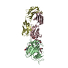
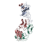
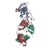
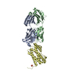
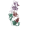
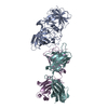
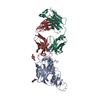
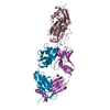
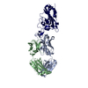
| Title | Crystal structure of SARS-CoV-2 spike RBD in complex with human monoclonal antibody AZD8895 | ||||||
 Components Components |
| ||||||
 Keywords Keywords | VIRAL PROTEIN/IMMUNE SYSTEM / SARS-CoV-2 / spike protein / receptor binding domain / human monoclonal antibody / VIRAL PROTEIN-IMMUNE SYSTEM complex | ||||||
| Function / homology |  Function and homology information Function and homology informationsymbiont-mediated disruption of host tissue / Maturation of spike protein / Translation of Structural Proteins / Virion Assembly and Release / host cell surface / host extracellular region / symbiont-mediated-mediated suppression of host tetherin activity / Induction of Cell-Cell Fusion / structural constituent of virion / positive regulation of viral entry into host cell ...symbiont-mediated disruption of host tissue / Maturation of spike protein / Translation of Structural Proteins / Virion Assembly and Release / host cell surface / host extracellular region / symbiont-mediated-mediated suppression of host tetherin activity / Induction of Cell-Cell Fusion / structural constituent of virion / positive regulation of viral entry into host cell / membrane fusion / host cell endoplasmic reticulum-Golgi intermediate compartment membrane / Attachment and Entry / entry receptor-mediated virion attachment to host cell / receptor-mediated virion attachment to host cell / host cell surface receptor binding / symbiont-mediated suppression of host innate immune response / endocytosis involved in viral entry into host cell / receptor ligand activity / fusion of virus membrane with host plasma membrane / fusion of virus membrane with host endosome membrane / viral envelope / symbiont entry into host cell / virion attachment to host cell / host cell plasma membrane / SARS-CoV-2 activates/modulates innate and adaptive immune responses / virion membrane / membrane / identical protein binding / plasma membrane Similarity search - Function | ||||||
| Biological species |  Homo sapiens (human) Homo sapiens (human)  Severe acute respiratory syndrome coronavirus 2 Severe acute respiratory syndrome coronavirus 2 | ||||||
| Method |  X-RAY DIFFRACTION / SYNCHROTRON / MOLECULAR REPLACEMENT / Resolution: 2.5 Å X-RAY DIFFRACTION / SYNCHROTRON / MOLECULAR REPLACEMENT / Resolution: 2.5 Å | ||||||
 Authors Authors | Dong, J. / Crowe, J.E. | ||||||
 Citation Citation | Journal: Nat Microbiol / Year: 2021 Title: Genetic and structural basis for SARS-CoV-2 variant neutralization by a two-antibody cocktail. Authors: Dong, J. / Zost, S.J. / Greaney, A.J. / Starr, T.N. / Dingens, A.S. / Chen, E.C. / Chen, R.E. / Case, J.B. / Sutton, R.E. / Gilchuk, P. / Rodriguez, J. / Armstrong, E. / Gainza, C. / Nargi, ...Authors: Dong, J. / Zost, S.J. / Greaney, A.J. / Starr, T.N. / Dingens, A.S. / Chen, E.C. / Chen, R.E. / Case, J.B. / Sutton, R.E. / Gilchuk, P. / Rodriguez, J. / Armstrong, E. / Gainza, C. / Nargi, R.S. / Binshtein, E. / Xie, X. / Zhang, X. / Shi, P.Y. / Logue, J. / Weston, S. / McGrath, M.E. / Frieman, M.B. / Brady, T. / Tuffy, K.M. / Bright, H. / Loo, Y.M. / McTamney, P.M. / Esser, M.T. / Carnahan, R.H. / Diamond, M.S. / Bloom, J.D. / Crowe Jr., J.E. | ||||||
| History |
|
- Structure visualization
| Structure viewer | Molecule: MolmilJmol/JSmol |
|---|
- Downloads & links
-Download
| PDBx/mmCIF format | 7l7d.cif.gz | 266.9 KB | Display | PDBx/mmCIF format |
|---|---|---|---|---|
| PDB format | pdb7l7d.ent.gz | 213.2 KB | Display | PDB format |
| PDBx/mmJSON format | 7l7d.json.gz | Tree view | PDBx/mmJSON format | |
| Others |  Other downloads Other downloads |
-Validation report
| Arichive directory | https://data.pdbj.org/pub/pdb/validation_reports/l7/7l7dftp://data.pdbj.org/pub/pdb/validation_reports/l7/7l7d | HTTPS FTP |
|---|
-Related structure data
| Related structure data | 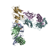 7l7eC  6xc2S S: Starting model for refinement C: citing same article ( |
|---|---|
| Similar structure data |

-Links
 PDBj
PDBj


- Assembly
| Deposited unit | 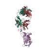
| ||||||||
|---|---|---|---|---|---|---|---|---|---|
| 1 |
| ||||||||
| Unit cell |
|
-Components
-Antibody , 2 types, 2 molecules HL
| #1: Antibody | Mass: 24100.277 Da / Num. of mol.: 1 Source method: isolated from a genetically manipulated source Source: (gene. exp.) Homo sapiens (human) / Production host: Homo sapiens (human) |
|---|---|
| #2: Antibody | Mass: 23644.217 Da / Num. of mol.: 1 Source method: isolated from a genetically manipulated source Source: (gene. exp.) Homo sapiens (human) / Production host: Homo sapiens (human) |
-Protein / Sugars , 2 types, 2 molecules E
| #3: Protein | Mass: 24229.189 Da / Num. of mol.: 1 / Fragment: receptor binding domain (UNP residues 330-529) Source method: isolated from a genetically manipulated source Source: (gene. exp.) Severe acute respiratory syndrome coronavirus 2Gene: S, 2 / Production host: Homo sapiens (human) / References: UniProt: P0DTC2 |
|---|---|
| #5: Sugar | ChemComp-NAG /  Type: D-saccharide, beta linking / Mass: 221.208 Da / Num. of mol.: 1 / Source method: isolated from a natural source / Formula: C8H15NO6 Type: D-saccharide, beta linking / Mass: 221.208 Da / Num. of mol.: 1 / Source method: isolated from a natural source / Formula: C8H15NO6 |
-Non-polymers , 2 types, 225 molecules 

| #4: Chemical | ChemComp-GOL /  Mass: 92.094 Da / Num. of mol.: 6 / Source method: obtained synthetically / Formula: C3H8O3 Mass: 92.094 Da / Num. of mol.: 6 / Source method: obtained synthetically / Formula: C3H8O3#6: Water | ChemComp-HOH / | Mass: 18.015 Da / Num. of mol.: 219 / Source method: isolated from a natural source / Formula: H2O |
|---|
-Details
| Has ligand of interest | N |
|---|---|
| Has protein modification | Y |
-Experimental details
-Experiment
| Experiment | Method: X-RAY DIFFRACTION / Number of used crystals: 1 |
|---|
- Sample preparation
| Crystal | Density Matthews: 2.63 Å3/Da / Density % sol: 53.19 % |
|---|---|
| Crystal grow | Temperature: 291 K / Method: vapor diffusion, sitting drop / Details: 16% PEG3350, 0.2 M Tris-HCl, pH 8.5 |
-Data collection
| Diffraction | Mean temperature: 100 K / Serial crystal experiment: N |
|---|---|
| Diffraction source | Source: SYNCHROTRON / Site: APS  / Beamline: 21-ID-F / Wavelength: 0.97872 Å / Beamline: 21-ID-F / Wavelength: 0.97872 Å |
| Detector | Type: RAYONIX MX-300 / Detector: CCD / Date: Sep 17, 2020 |
| Radiation | Monochromator: diamond(111) / Protocol: SINGLE WAVELENGTH / Monochromatic (M) / Laue (L): M / Scattering type: x-ray |
| Radiation wavelength | Wavelength: 0.97872 Å / Relative weight: 1 |
| Reflection | Resolution: 2.5→43.98 Å / Num. obs: 24896 / % possible obs: 99.9 % / Redundancy: 3.7 % / Rmerge(I) obs: 0.046 / Net I/σ(I): 19.2 |
| Reflection shell | Resolution: 2.5→2.63 Å / Rmerge(I) obs: 0.245 / Num. unique obs: 3598 |
- Processing
| Software |
| ||||||||||||||||||||||||||||||||||||||||||||||||||||||||||||||||||||||
|---|---|---|---|---|---|---|---|---|---|---|---|---|---|---|---|---|---|---|---|---|---|---|---|---|---|---|---|---|---|---|---|---|---|---|---|---|---|---|---|---|---|---|---|---|---|---|---|---|---|---|---|---|---|---|---|---|---|---|---|---|---|---|---|---|---|---|---|---|---|---|---|
| Refinement | Method to determine structure: MOLECULAR REPLACEMENT Starting model: PDB entry 6XC2 Resolution: 2.5→43.98 Å / SU ML: 0.29 / Cross valid method: FREE R-VALUE / σ(F): 1.35 / Phase error: 24.09 / Stereochemistry target values: ML
| ||||||||||||||||||||||||||||||||||||||||||||||||||||||||||||||||||||||
| Solvent computation | Shrinkage radii: 0.9 Å / VDW probe radii: 1.11 Å / Solvent model: FLAT BULK SOLVENT MODEL | ||||||||||||||||||||||||||||||||||||||||||||||||||||||||||||||||||||||
| Refinement step | Cycle: LAST / Resolution: 2.5→43.98 Å
| ||||||||||||||||||||||||||||||||||||||||||||||||||||||||||||||||||||||
| Refine LS restraints |
| ||||||||||||||||||||||||||||||||||||||||||||||||||||||||||||||||||||||
| LS refinement shell |
| ||||||||||||||||||||||||||||||||||||||||||||||||||||||||||||||||||||||
| Refinement TLS params. | Method: refined / Origin x: -17.4857 Å / Origin y: 15.6466 Å / Origin z: 53.4486 Å
| ||||||||||||||||||||||||||||||||||||||||||||||||||||||||||||||||||||||
| Refinement TLS group | Selection details: all |
