 Movie
Movie Controller

[English] 日本語
 Yorodumi
- PDB-7l2c: Crystallographic structure of neutralizing antibody 2-51 in compl... -
+ Open data

- Basic information
| Entry | Database: PDB / ID: 7l2c | ||||||
|---|---|---|---|---|---|---|---|
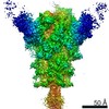
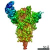
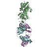
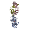
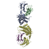
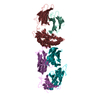
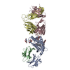
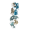
| Title | Crystallographic structure of neutralizing antibody 2-51 in complex with SARS-CoV-2 spike N-terminal domain (NTD) | ||||||
 Components Components |
| ||||||
 Keywords Keywords | IMMUNE SYSTEM/Viral Protein / COVID-19 / SARS-CoV-2 / Viral protein / Spike glycoprotein / N-terminal domain / NTD / Neutralizing antibody / 2-51 / Fab / IMMUNE SYSTEM / IMMUNE SYSTEM-Viral Protein complex | ||||||
| Function / homology |  Function and homology information Function and homology informationsymbiont-mediated disruption of host tissue / Maturation of spike protein / Translation of Structural Proteins / Virion Assembly and Release / host cell surface / host extracellular space / viral translation / symbiont-mediated-mediated suppression of host tetherin activity / Induction of Cell-Cell Fusion / structural constituent of virion ...symbiont-mediated disruption of host tissue / Maturation of spike protein / Translation of Structural Proteins / Virion Assembly and Release / host cell surface / host extracellular space / viral translation / symbiont-mediated-mediated suppression of host tetherin activity / Induction of Cell-Cell Fusion / structural constituent of virion / membrane fusion / entry receptor-mediated virion attachment to host cell / Attachment and Entry / host cell endoplasmic reticulum-Golgi intermediate compartment membrane / positive regulation of viral entry into host cell / receptor-mediated virion attachment to host cell / host cell surface receptor binding / symbiont-mediated suppression of host innate immune response / receptor ligand activity / endocytosis involved in viral entry into host cell / fusion of virus membrane with host plasma membrane / fusion of virus membrane with host endosome membrane / viral envelope / symbiont entry into host cell / virion attachment to host cell / SARS-CoV-2 activates/modulates innate and adaptive immune responses / host cell plasma membrane / virion membrane / identical protein binding / membrane / plasma membrane Similarity search - Function | ||||||
| Biological species |   Severe acute respiratory syndrome coronavirus 2 Severe acute respiratory syndrome coronavirus 2 Homo sapiens (human) Homo sapiens (human) | ||||||
| Method |  X-RAY DIFFRACTION / SYNCHROTRON / MOLECULAR REPLACEMENT / Resolution: 3.65 Å X-RAY DIFFRACTION / SYNCHROTRON / MOLECULAR REPLACEMENT / Resolution: 3.65 Å | ||||||
 Authors Authors | Cerutti, G. / Reddem, E.R. / Shapiro, L. | ||||||
| Funding support |  China, 1items China, 1items
| ||||||
 Citation Citation | Journal: Cell Host Microbe / Year: 2021 Title: Potent SARS-CoV-2 neutralizing antibodies directed against spike N-terminal domain target a single supersite. Authors: Gabriele Cerutti / Yicheng Guo / Tongqing Zhou / Jason Gorman / Myungjin Lee / Micah Rapp / Eswar R Reddem / Jian Yu / Fabiana Bahna / Jude Bimela / Yaoxing Huang / Phinikoula S Katsamba / ...Authors: Gabriele Cerutti / Yicheng Guo / Tongqing Zhou / Jason Gorman / Myungjin Lee / Micah Rapp / Eswar R Reddem / Jian Yu / Fabiana Bahna / Jude Bimela / Yaoxing Huang / Phinikoula S Katsamba / Lihong Liu / Manoj S Nair / Reda Rawi / Adam S Olia / Pengfei Wang / Baoshan Zhang / Gwo-Yu Chuang / David D Ho / Zizhang Sheng / Peter D Kwong / Lawrence Shapiro /  Abstract: Numerous antibodies that neutralize SARS-CoV-2 have been identified, and these generally target either the receptor-binding domain (RBD) or the N-terminal domain (NTD) of the viral spike. While RBD- ...Numerous antibodies that neutralize SARS-CoV-2 have been identified, and these generally target either the receptor-binding domain (RBD) or the N-terminal domain (NTD) of the viral spike. While RBD-directed antibodies have been extensively studied, far less is known about NTD-directed antibodies. Here, we report cryo-EM and crystal structures for seven potent NTD-directed neutralizing antibodies in complex with spike or isolated NTD. These structures defined several antibody classes, with at least one observed in multiple convalescent donors. The structures revealed that all seven antibodies target a common surface, bordered by glycans N17, N74, N122, and N149. This site-formed primarily by a mobile β-hairpin and several flexible loops-was highly electropositive, located at the periphery of the spike, and the largest glycan-free surface of NTD facing away from the viral membrane. Thus, in contrast to neutralizing RBD-directed antibodies that recognize multiple non-overlapping epitopes, potent NTD-directed neutralizing antibodies appear to target a single supersite. | ||||||
| History |
|
- Structure visualization
| Structure viewer | Molecule: MolmilJmol/JSmol |
|---|
- Downloads & links
-Download
| PDBx/mmCIF format | 7l2c.cif.gz | 300.7 KB | Display | PDBx/mmCIF format |
|---|---|---|---|---|
| PDB format | pdb7l2c.ent.gz | 238.4 KB | Display | PDB format |
| PDBx/mmJSON format | 7l2c.json.gz | Tree view | PDBx/mmJSON format | |
| Others |  Other downloads Other downloads |
-Validation report
| Summary document | 7l2c_validation.pdf.gz | 13.6 MB | Display | wwPDB validaton report |
|---|---|---|---|---|
| Full document | 7l2c_full_validation.pdf.gz | 13.6 MB | Display | |
| Data in XML | 7l2c_validation.xml.gz | 52.6 KB | Display | |
| Data in CIF | 7l2c_validation.cif.gz | 71.9 KB | Display | |
| Arichive directory | https://data.pdbj.org/pub/pdb/validation_reports/l2/7l2cftp://data.pdbj.org/pub/pdb/validation_reports/l2/7l2c | HTTPS FTP |
-Related structure data
| Related structure data |  7l2dC  7l2eC  7l2fC  7lqvC  7lqwC  7c2lS S: Starting model for refinement C: citing same article ( |
|---|---|
| Similar structure data |


-Links
 PDBj
PDBj


- Assembly
| Deposited unit | 
| ||||||||
|---|---|---|---|---|---|---|---|---|---|
| 1 | 
| ||||||||
| 2 | 
| ||||||||
| Unit cell |
|
-Components
-Protein , 1 types, 2 molecules AB
| #1: Protein | Mass: 38861.996 Da / Num. of mol.: 2 Source method: isolated from a genetically manipulated source Source: (gene. exp.) Severe acute respiratory syndrome coronavirus 2Gene: S, 2 / Production host: Homo sapiens (human) / References: UniProt: P0DTC2 |
|---|
-Antibody , 2 types, 4 molecules HCLD
| #2: Antibody | Mass: 24369.305 Da / Num. of mol.: 2 Source method: isolated from a genetically manipulated source Source: (gene. exp.) Homo sapiens (human) / Production host: Homo sapiens (human)#3: Antibody | Mass: 22591.912 Da / Num. of mol.: 2 Source method: isolated from a genetically manipulated source Source: (gene. exp.) Homo sapiens (human) / Production host: Homo sapiens (human) |
|---|
-Sugars , 2 types, 14 molecules 
| #4: Polysaccharide | 2-acetamido-2-deoxy-beta-D-glucopyranose-(1-4)-2-acetamido-2-deoxy-beta-D-glucopyranose Source method: isolated from a genetically manipulated source |
|---|---|
| #5: Sugar | ChemComp-NAG /  Type: D-saccharide, beta linking / Mass: 221.208 Da / Num. of mol.: 13 / Source method: obtained synthetically / Formula: C8H15NO6 / Feature type: SUBJECT OF INVESTIGATION Type: D-saccharide, beta linking / Mass: 221.208 Da / Num. of mol.: 13 / Source method: obtained synthetically / Formula: C8H15NO6 / Feature type: SUBJECT OF INVESTIGATION |
-Non-polymers , 5 types, 91 molecules 


| #6: Chemical | ChemComp-CA /  Mass: 40.078 Da / Num. of mol.: 39 / Source method: obtained synthetically / Formula: Ca / Feature type: SUBJECT OF INVESTIGATION Mass: 40.078 Da / Num. of mol.: 39 / Source method: obtained synthetically / Formula: Ca / Feature type: SUBJECT OF INVESTIGATION#7: Chemical | ChemComp-ACT /  Mass: 59.044 Da / Num. of mol.: 9 / Source method: obtained synthetically / Formula: C2H3O2 / Feature type: SUBJECT OF INVESTIGATION Mass: 59.044 Da / Num. of mol.: 9 / Source method: obtained synthetically / Formula: C2H3O2 / Feature type: SUBJECT OF INVESTIGATION#8: Chemical | ChemComp-CAC / |  Mass: 136.989 Da / Num. of mol.: 1 / Source method: obtained synthetically / Formula: C2H6AsO2 / Feature type: SUBJECT OF INVESTIGATION Mass: 136.989 Da / Num. of mol.: 1 / Source method: obtained synthetically / Formula: C2H6AsO2 / Feature type: SUBJECT OF INVESTIGATION#9: Chemical | ChemComp-PGE / |  Mass: 150.173 Da / Num. of mol.: 1 / Source method: isolated from a natural source / Formula: C6H14O4 / Feature type: SUBJECT OF INVESTIGATION Mass: 150.173 Da / Num. of mol.: 1 / Source method: isolated from a natural source / Formula: C6H14O4 / Feature type: SUBJECT OF INVESTIGATION#10: Water | ChemComp-HOH / | Mass: 18.015 Da / Num. of mol.: 41 / Source method: isolated from a natural source / Formula: H2O |
|---|
-Details
| Has ligand of interest | Y |
|---|---|
| Has protein modification | Y |
-Experimental details
-Experiment
| Experiment | Method: X-RAY DIFFRACTION / Number of used crystals: 1 |
|---|
- Sample preparation
| Crystal | Density Matthews: 3.05 Å3/Da / Density % sol: 63.38 % |
|---|---|
| Crystal grow | Temperature: 298 K / Method: vapor diffusion, sitting drop / pH: 6.5 Details: 0.16 M Calcium Acetate, 0.08 M Sodium Cacodylate, 14.4% PEG 8000, 20% Glycerol |
-Data collection
| Diffraction | Mean temperature: 100 K / Serial crystal experiment: N |
|---|---|
| Diffraction source | Source: SYNCHROTRON / Site: APS / Beamline: 24-ID-C / Wavelength: 0.979 Å |
| Detector | Type: DECTRIS EIGER X 16M / Detector: PIXEL / Date: Oct 14, 2020 |
| Radiation | Protocol: SINGLE WAVELENGTH / Monochromatic (M) / Laue (L): M / Scattering type: x-ray |
| Radiation wavelength | Wavelength: 0.979 Å / Relative weight: 1 |
| Reflection | Resolution: 3.44→88.03 Å / Num. obs: 25964 / % possible obs: 93.26 % / Redundancy: 1.9 % / CC1/2: 0.883 / Net I/σ(I): 2.57 |
| Reflection shell | Resolution: 3.44→3.65 Å / Num. unique obs: 5916 / CC1/2: 0.44 |
- Processing
| Software |
| |||||||||||||||||||||||||||||||||||||||||||||||||||||||||||||||
|---|---|---|---|---|---|---|---|---|---|---|---|---|---|---|---|---|---|---|---|---|---|---|---|---|---|---|---|---|---|---|---|---|---|---|---|---|---|---|---|---|---|---|---|---|---|---|---|---|---|---|---|---|---|---|---|---|---|---|---|---|---|---|---|---|
| Refinement | Method to determine structure: MOLECULAR REPLACEMENT Starting model: 7C2L Resolution: 3.65→88.03 Å / SU ML: 0.46 / Cross valid method: FREE R-VALUE / σ(F): 1.35 / Phase error: 27.12 / Stereochemistry target values: ML
| |||||||||||||||||||||||||||||||||||||||||||||||||||||||||||||||
| Solvent computation | Shrinkage radii: 0.9 Å / VDW probe radii: 1.11 Å / Solvent model: FLAT BULK SOLVENT MODEL | |||||||||||||||||||||||||||||||||||||||||||||||||||||||||||||||
| Refinement step | Cycle: LAST / Resolution: 3.65→88.03 Å
| |||||||||||||||||||||||||||||||||||||||||||||||||||||||||||||||
| Refine LS restraints |
| |||||||||||||||||||||||||||||||||||||||||||||||||||||||||||||||
| LS refinement shell |
|
