 Movie
Movie Controller

[English] 日本語
 Yorodumi
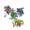
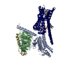
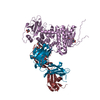
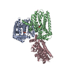
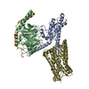
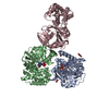
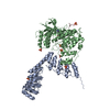
- PDB-7l0r: Structure of NTS-NTSR1-Gi complex in lipid nanodisc, noncanonical... -
+ Open data

- Basic information
| Entry | Database: PDB / ID: 7l0r | ||||||
|---|---|---|---|---|---|---|---|
| Title | Structure of NTS-NTSR1-Gi complex in lipid nanodisc, noncanonical state, without AHD | ||||||
 Components Components |
| ||||||
 Keywords Keywords | SIGNALING PROTEIN / GPCR / NTSR1 / NTS / G protein / Nanodisc | ||||||
| Function / homology |  Function and homology information Function and homology informationregulation of locomotion involved in locomotory behavior / Peptide ligand-binding receptors / neuropeptide receptor binding / eye photoreceptor cell development / G protein-coupled neurotensin receptor activity / inositol phosphate catabolic process / symmetric synapse / positive regulation of locomotion / regulation of inositol trisphosphate biosynthetic process / positive regulation of cation channel activity ...regulation of locomotion involved in locomotory behavior / Peptide ligand-binding receptors / neuropeptide receptor binding / eye photoreceptor cell development / G protein-coupled neurotensin receptor activity / inositol phosphate catabolic process / symmetric synapse / positive regulation of locomotion / regulation of inositol trisphosphate biosynthetic process / positive regulation of cation channel activity / response to antipsychotic drug / positive regulation of gamma-aminobutyric acid secretion / D-aspartate import across plasma membrane / hyperosmotic response / neuron spine / neuropeptide hormone activity / positive regulation of arachidonate secretion / vocalization behavior / L-glutamate import across plasma membrane / regulation of behavioral fear response / digestive tract development / cAMP biosynthetic process / regulation of respiratory gaseous exchange / negative regulation of systemic arterial blood pressure / G alpha (q) signalling events / positive regulation of inhibitory postsynaptic potential / negative regulation of release of sequestered calcium ion into cytosol / cellular response to lithium ion / response to mineralocorticoid / response to food / positive regulation of glutamate secretion / response to corticosterone / regulation of membrane depolarization / response to lipid / temperature homeostasis / positive regulation of inositol phosphate biosynthetic process / response to stress / detection of temperature stimulus involved in sensory perception of pain / associative learning / phototransduction / cellular response to dexamethasone stimulus / response to axon injury / conditioned place preference / transport vesicle / adult locomotory behavior / regulation of eating behavior / cardiac muscle cell apoptotic process / adenylate cyclase inhibitor activity / photoreceptor inner segment / positive regulation of protein localization to cell cortex / T cell migration / positive regulation of relaxation of smooth muscle / Adenylate cyclase inhibitory pathway / D2 dopamine receptor binding / adenylate cyclase-inhibiting serotonin receptor signaling pathway / G protein-coupled serotonin receptor binding / blood vessel diameter maintenance / axon terminus / response to amphetamine / positive regulation of release of sequestered calcium ion into cytosol / liver development / cellular response to forskolin / mast cell degranulation / regulation of mitotic spindle organization / learning / dendritic shaft / chemokine-mediated signaling pathway / response to cocaine / Regulation of insulin secretion / neuropeptide signaling pathway / response to prostaglandin E / cellular response to nerve growth factor stimulus / visual learning / positive regulation of cholesterol biosynthetic process / G protein-coupled receptor binding / response to peptide hormone / cytoplasmic side of plasma membrane / G-protein beta/gamma-subunit complex binding / adenylate cyclase-modulating G protein-coupled receptor signaling pathway / adenylate cyclase-inhibiting G protein-coupled receptor signaling pathway / Olfactory Signaling Pathway / terminal bouton / Activation of the phototransduction cascade / G protein-coupled acetylcholine receptor signaling pathway / G beta:gamma signalling through PLC beta / Presynaptic function of Kainate receptors / Thromboxane signalling through TP receptor / Activation of G protein gated Potassium channels / Inhibition of voltage gated Ca2+ channels via Gbeta/gamma subunits / G-protein activation / Glucagon signaling in metabolic regulation / G beta:gamma signalling through CDC42 / Prostacyclin signalling through prostacyclin receptor / intracellular protein localization / Synthesis, secretion, and inactivation of Glucagon-like Peptide-1 (GLP-1) / response to estradiol / G beta:gamma signalling through BTK / photoreceptor disc membrane / GDP binding / ADP signalling through P2Y purinoceptor 12 Similarity search - Function | ||||||
| Biological species |   Homo sapiens (human) Homo sapiens (human) | ||||||
| Method | ELECTRON MICROSCOPY / single particle reconstruction / cryo EM / Resolution: 4.2 Å | ||||||
 Authors Authors | Zhang, M. / Gui, M. / Wang, Z. / Gorgulla, C. / Yu, J.J. / Wu, H. / Sun, Z. / Klenk, C. / Merklinger, L. / Morstein, L. ...Zhang, M. / Gui, M. / Wang, Z. / Gorgulla, C. / Yu, J.J. / Wu, H. / Sun, Z. / Klenk, C. / Merklinger, L. / Morstein, L. / Hagn, F. / Pluckthun, A. / Brown, A. / Nasr, M.L. / Wagner, G. | ||||||
 Citation Citation | Journal: Nat Struct Mol Biol / Year: 2021 Title: Cryo-EM structure of an activated GPCR-G protein complex in lipid nanodiscs. Authors: Meng Zhang / Miao Gui / Zi-Fu Wang / Christoph Gorgulla / James J Yu / Hao Wu / Zhen-Yu J Sun / Christoph Klenk / Lisa Merklinger / Lena Morstein / Franz Hagn / Andreas Plückthun / Alan ...Authors: Meng Zhang / Miao Gui / Zi-Fu Wang / Christoph Gorgulla / James J Yu / Hao Wu / Zhen-Yu J Sun / Christoph Klenk / Lisa Merklinger / Lena Morstein / Franz Hagn / Andreas Plückthun / Alan Brown / Mahmoud L Nasr / Gerhard Wagner /    Abstract: G-protein-coupled receptors (GPCRs) are the largest superfamily of transmembrane proteins and the targets of over 30% of currently marketed pharmaceuticals. Although several structures have been ...G-protein-coupled receptors (GPCRs) are the largest superfamily of transmembrane proteins and the targets of over 30% of currently marketed pharmaceuticals. Although several structures have been solved for GPCR-G protein complexes, few are in a lipid membrane environment. Here, we report cryo-EM structures of complexes of neurotensin, neurotensin receptor 1 and Gαβγ in two conformational states, resolved to resolutions of 4.1 and 4.2 Å. The structures, determined in a lipid bilayer without any stabilizing antibodies or nanobodies, reveal an extended network of protein-protein interactions at the GPCR-G protein interface as compared to structures obtained in detergent micelles. The findings show that the lipid membrane modulates the structure and dynamics of complex formation and provide a molecular explanation for the stronger interaction between GPCRs and G proteins in lipid bilayers. We propose an allosteric mechanism for GDP release, providing new insights into the activation of G proteins for downstream signaling. | ||||||
| History |
|
- Structure visualization
| Movie |
Movie viewer |
|---|---|
| Structure viewer | Molecule: MolmilJmol/JSmol |
- Downloads & links
-Download
| PDBx/mmCIF format | 7l0r.cif.gz | 191.6 KB | Display | PDBx/mmCIF format |
|---|---|---|---|---|
| PDB format | pdb7l0r.ent.gz | 144.6 KB | Display | PDB format |
| PDBx/mmJSON format | 7l0r.json.gz | Tree view | PDBx/mmJSON format | |
| Others |  Other downloads Other downloads |
-Validation report
| Arichive directory | https://data.pdbj.org/pub/pdb/validation_reports/l0/7l0rftp://data.pdbj.org/pub/pdb/validation_reports/l0/7l0r | HTTPS FTP |
|---|
-Related structure data
| Related structure data |  23101MC  7l0pC  7l0qC  7l0sC C: citing same article ( M: map data used to model this data |
|---|---|
| Similar structure data |


-Links
 PDBj
PDBj


- Assembly
| Deposited unit | 
|
|---|---|
| 1 |
|
-Components
| #1: Protein | Mass: 37483.016 Da / Num. of mol.: 1 Mutation: A86L, H103D, H105Y, A161V, R213L, V234L, I253A, H305R, F358V, S362A, del273-290 Source method: isolated from a genetically manipulated source Source: (gene. exp.)  |
|---|---|
| #2: Protein/peptide | Mass: 1087.277 Da / Num. of mol.: 1 / Fragment: residues 157-162 Source method: isolated from a genetically manipulated source Source: (gene. exp.) |
| #3: Protein | Mass: 40415.031 Da / Num. of mol.: 1 Source method: isolated from a genetically manipulated source Source: (gene. exp.) Homo sapiens (human) / Gene: GNAI1 / Production host:   Spodoptera frugiperda (fall armyworm) / References: UniProt: P63096 Spodoptera frugiperda (fall armyworm) / References: UniProt: P63096 |
| #4: Protein | Mass: 39983.727 Da / Num. of mol.: 1 Source method: isolated from a genetically manipulated source Source: (gene. exp.) Homo sapiens (human) / Gene: GNB1 / Production host: Spodoptera frugiperda (fall armyworm) / References: UniProt: P62873 |
| #5: Protein | Mass: 9593.011 Da / Num. of mol.: 1 Source method: isolated from a genetically manipulated source Source: (gene. exp.) Homo sapiens (human) / Gene: GNGT1 / Production host: Spodoptera frugiperda (fall armyworm) / References: UniProt: P63211 |
| Has protein modification | Y |
-Experimental details
-Experiment
| Experiment | Method: ELECTRON MICROSCOPY |
|---|---|
| EM experiment | Aggregation state: PARTICLE / 3D reconstruction method: single particle reconstruction |
- Sample preparation
| Component |
| ||||||||||||||||||||||||||||||||||||||||||
|---|---|---|---|---|---|---|---|---|---|---|---|---|---|---|---|---|---|---|---|---|---|---|---|---|---|---|---|---|---|---|---|---|---|---|---|---|---|---|---|---|---|---|---|
| Molecular weight |
| ||||||||||||||||||||||||||||||||||||||||||
| Source (natural) |
| ||||||||||||||||||||||||||||||||||||||||||
| Source (recombinant) |
| ||||||||||||||||||||||||||||||||||||||||||
| Buffer solution | pH: 6.9 | ||||||||||||||||||||||||||||||||||||||||||
| Specimen | Embedding applied: NO / Shadowing applied: NO / Staining applied: NO / Vitrification applied: YES | ||||||||||||||||||||||||||||||||||||||||||
| Vitrification | Cryogen name: ETHANE |
- Electron microscopy imaging
| Experimental equipment |  Model: Titan Krios / Image courtesy: FEI Company |
|---|---|
| Microscopy | Model: FEI TITAN KRIOS |
| Electron gun | Electron source:  FIELD EMISSION GUN / Accelerating voltage: 300 kV / Illumination mode: FLOOD BEAM FIELD EMISSION GUN / Accelerating voltage: 300 kV / Illumination mode: FLOOD BEAM |
| Electron lens | Mode: BRIGHT FIELD |
| Image recording | Electron dose: 57 e/Å2 / Film or detector model: GATAN K3 BIOQUANTUM (6k x 4k) |
- Processing
| Software | Name: PHENIX / Version: dev_3318: / Classification: refinement | ||||||||||||||||||||||||||||||||||||||||
|---|---|---|---|---|---|---|---|---|---|---|---|---|---|---|---|---|---|---|---|---|---|---|---|---|---|---|---|---|---|---|---|---|---|---|---|---|---|---|---|---|---|
| EM software |
| ||||||||||||||||||||||||||||||||||||||||
| CTF correction | Type: PHASE FLIPPING AND AMPLITUDE CORRECTION | ||||||||||||||||||||||||||||||||||||||||
| Particle selection | Num. of particles selected: 4367542 | ||||||||||||||||||||||||||||||||||||||||
| Symmetry | Point symmetry: C1 (asymmetric) | ||||||||||||||||||||||||||||||||||||||||
| 3D reconstruction | Resolution: 4.2 Å / Resolution method: FSC 0.143 CUT-OFF / Num. of particles: 324002 / Symmetry type: POINT | ||||||||||||||||||||||||||||||||||||||||
| Atomic model building | Protocol: FLEXIBLE FIT / Space: REAL | ||||||||||||||||||||||||||||||||||||||||
| Refine LS restraints |
|
