 Movie
Movie Controller

[English] 日本語
 Yorodumi
- EMDB-23101: Structure of NTS-NTSR1-Gi complex in lipid nanodisc, noncanonical... -
+ Open data

- Basic information
| Entry | Database: EMDB / ID: EMD-23101 | |||||||||
|---|---|---|---|---|---|---|---|---|---|---|
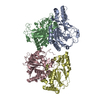
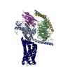
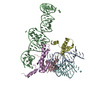
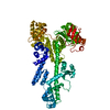
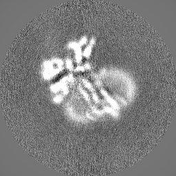
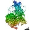
| Title | Structure of NTS-NTSR1-Gi complex in lipid nanodisc, noncanonical state, AHD and nanodisc mask out | |||||||||
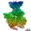
 Map data Map data | ||||||||||
 Sample Sample |
| |||||||||
 Keywords Keywords | GPCR / NTSR1 / NTS / G protein / Nanodisc / SIGNALING PROTEIN | |||||||||
| Function / homology |  Function and homology information Function and homology informationregulation of locomotion involved in locomotory behavior / Peptide ligand-binding receptors / neuropeptide receptor binding / G protein-coupled neurotensin receptor activity / inositol phosphate catabolic process / symmetric synapse / positive regulation of locomotion / eye photoreceptor cell development / regulation of inositol trisphosphate biosynthetic process / response to antipsychotic drug ...regulation of locomotion involved in locomotory behavior / Peptide ligand-binding receptors / neuropeptide receptor binding / G protein-coupled neurotensin receptor activity / inositol phosphate catabolic process / symmetric synapse / positive regulation of locomotion / eye photoreceptor cell development / regulation of inositol trisphosphate biosynthetic process / response to antipsychotic drug / positive regulation of gamma-aminobutyric acid secretion / D-aspartate import across plasma membrane / neuron spine / neuropeptide hormone activity / positive regulation of arachidonate secretion / L-glutamate import across plasma membrane / vocalization behavior / regulation of behavioral fear response / cAMP biosynthetic process / regulation of respiratory gaseous exchange / G alpha (q) signalling events / hyperosmotic response / digestive tract development / positive regulation of inhibitory postsynaptic potential / negative regulation of systemic arterial blood pressure / negative regulation of release of sequestered calcium ion into cytosol / cellular response to lithium ion / response to mineralocorticoid / response to corticosterone / positive regulation of glutamate secretion / response to food / regulation of membrane depolarization / response to lipid / temperature homeostasis / positive regulation of inositol phosphate biosynthetic process / detection of temperature stimulus involved in sensory perception of pain / response to stress / phototransduction / associative learning / cellular response to dexamethasone stimulus / response to axon injury / conditioned place preference / transport vesicle / cardiac muscle cell apoptotic process / adenylate cyclase inhibitor activity / positive regulation of protein localization to cell cortex / photoreceptor inner segment / T cell migration / positive regulation of relaxation of smooth muscle / Adenylate cyclase inhibitory pathway / D2 dopamine receptor binding / adenylate cyclase-inhibiting serotonin receptor signaling pathway / G protein-coupled serotonin receptor binding / axon terminus / blood vessel diameter maintenance / cellular response to forskolin / positive regulation of release of sequestered calcium ion into cytosol / regulation of mitotic spindle organization / chemokine-mediated signaling pathway / dendritic shaft / learning / response to amphetamine / adult locomotory behavior / response to cocaine / liver development / Regulation of insulin secretion / neuropeptide signaling pathway / response to prostaglandin E / cellular response to nerve growth factor stimulus / positive regulation of cholesterol biosynthetic process / negative regulation of insulin secretion / visual learning / G protein-coupled receptor binding / response to peptide hormone / cytoplasmic side of plasma membrane / centriolar satellite / G-protein beta/gamma-subunit complex binding / adenylate cyclase-modulating G protein-coupled receptor signaling pathway / adenylate cyclase-inhibiting G protein-coupled receptor signaling pathway / Olfactory Signaling Pathway / Activation of the phototransduction cascade / terminal bouton / G protein-coupled acetylcholine receptor signaling pathway / G beta:gamma signalling through PLC beta / Presynaptic function of Kainate receptors / Thromboxane signalling through TP receptor / Activation of G protein gated Potassium channels / Inhibition of voltage gated Ca2+ channels via Gbeta/gamma subunits / G-protein activation / Glucagon signaling in metabolic regulation / Prostacyclin signalling through prostacyclin receptor / G beta:gamma signalling through CDC42 / Synthesis, secretion, and inactivation of Glucagon-like Peptide-1 (GLP-1) / G beta:gamma signalling through BTK / photoreceptor disc membrane / ADP signalling through P2Y purinoceptor 12 / intracellular protein localization / response to estradiol / Glucagon-type ligand receptors / Sensory perception of sweet, bitter, and umami (glutamate) taste Similarity search - Function | |||||||||
| Biological species |   Homo sapiens (human) Homo sapiens (human) | |||||||||
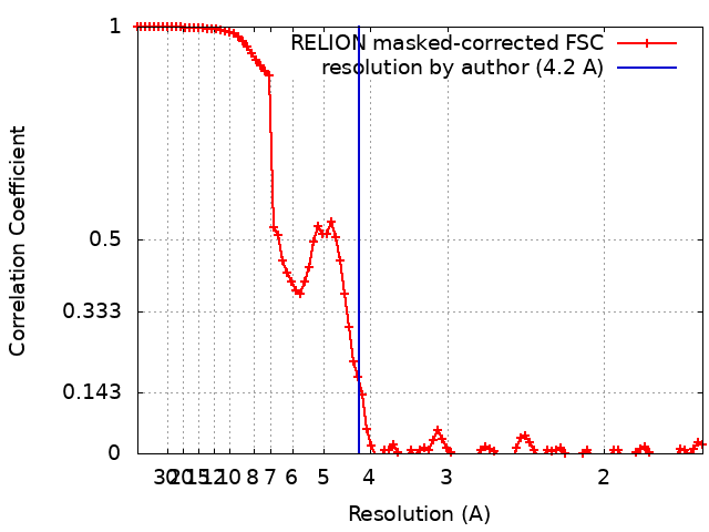
| Method | single particle reconstruction / cryo EM / Resolution: 4.2 Å | |||||||||
 Authors Authors | Zhang M / Gui M | |||||||||
 Citation Citation | Journal: Nat Struct Mol Biol / Year: 2021 Title: Cryo-EM structure of an activated GPCR-G protein complex in lipid nanodiscs. Authors: Meng Zhang / Miao Gui / Zi-Fu Wang / Christoph Gorgulla / James J Yu / Hao Wu / Zhen-Yu J Sun / Christoph Klenk / Lisa Merklinger / Lena Morstein / Franz Hagn / Andreas Plückthun / Alan ...Authors: Meng Zhang / Miao Gui / Zi-Fu Wang / Christoph Gorgulla / James J Yu / Hao Wu / Zhen-Yu J Sun / Christoph Klenk / Lisa Merklinger / Lena Morstein / Franz Hagn / Andreas Plückthun / Alan Brown / Mahmoud L Nasr / Gerhard Wagner /    Abstract: G-protein-coupled receptors (GPCRs) are the largest superfamily of transmembrane proteins and the targets of over 30% of currently marketed pharmaceuticals. Although several structures have been ...G-protein-coupled receptors (GPCRs) are the largest superfamily of transmembrane proteins and the targets of over 30% of currently marketed pharmaceuticals. Although several structures have been solved for GPCR-G protein complexes, few are in a lipid membrane environment. Here, we report cryo-EM structures of complexes of neurotensin, neurotensin receptor 1 and Gαβγ in two conformational states, resolved to resolutions of 4.1 and 4.2 Å. The structures, determined in a lipid bilayer without any stabilizing antibodies or nanobodies, reveal an extended network of protein-protein interactions at the GPCR-G protein interface as compared to structures obtained in detergent micelles. The findings show that the lipid membrane modulates the structure and dynamics of complex formation and provide a molecular explanation for the stronger interaction between GPCRs and G proteins in lipid bilayers. We propose an allosteric mechanism for GDP release, providing new insights into the activation of G proteins for downstream signaling. | |||||||||
| History |
|
- Structure visualization
| Movie |
Movie viewer |
|---|---|
| Structure viewer | EM map: SurfViewMolmilJmol/JSmol |
| Supplemental images |

- Downloads & links
-EMDB archive
| Map data | emd_23101.map.gz | 58.9 MB | EMDB map data format | |
|---|---|---|---|---|
| Header (meta data) | emd-23101-v30.xmlemd-23101.xml | 22.5 KB 22.5 KB | Display Display | EMDB header |
| FSC (resolution estimation) | emd_23101_fsc.xml | 9.2 KB | Display | FSC data file |
| Images | 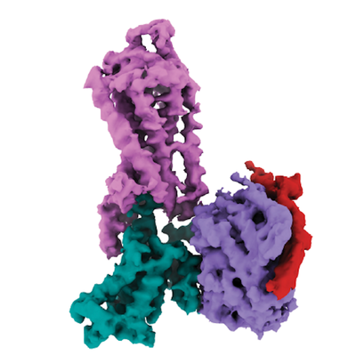 emd_23101.png emd_23101.png | 110 KB | ||
| Masks | emd_23101_msk_1.map | 64 MB | Mask map | |
| Filedesc metadata | emd-23101.cif.gz | 6.4 KB | ||
| Others | emd_23101_half_map_1.map.gzemd_23101_half_map_2.map.gz | 49.9 MB 49.9 MB | ||
| Archive directory |  http://ftp.pdbj.org/pub/emdb/structures/EMD-23101ftp://ftp.pdbj.org/pub/emdb/structures/EMD-23101 http://ftp.pdbj.org/pub/emdb/structures/EMD-23101ftp://ftp.pdbj.org/pub/emdb/structures/EMD-23101 | HTTPS FTP |
-Related structure data
| Related structure data |  7l0rMC  7l0pC  7l0qC  7l0sC C: citing same article ( M: atomic model generated by this map |
|---|---|
| Similar structure data |


-Links
| EMDB pages | EMDB (EBI/PDBe) / EMDataResource |
|---|---|
| Related items in Molecule of the Month |


-Map
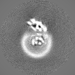
| File | Download / File: emd_23101.map.gz / Format: CCP4 / Size: 64 MB / Type: IMAGE STORED AS FLOATING POINT NUMBER (4 BYTES) | ||||||||||||||||||||||||||||||||||||||||||||||||||||||||||||
|---|---|---|---|---|---|---|---|---|---|---|---|---|---|---|---|---|---|---|---|---|---|---|---|---|---|---|---|---|---|---|---|---|---|---|---|---|---|---|---|---|---|---|---|---|---|---|---|---|---|---|---|---|---|---|---|---|---|---|---|---|---|
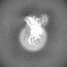
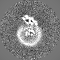
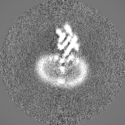
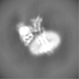
| Projections & slices | Image control
Images are generated by Spider. | ||||||||||||||||||||||||||||||||||||||||||||||||||||||||||||
| Voxel size | X=Y=Z: 0.825 Å | ||||||||||||||||||||||||||||||||||||||||||||||||||||||||||||
| Density |
| ||||||||||||||||||||||||||||||||||||||||||||||||||||||||||||
| Symmetry | Space group: 1 | ||||||||||||||||||||||||||||||||||||||||||||||||||||||||||||
| Details | EMDB XML:
CCP4 map header:
| ||||||||||||||||||||||||||||||||||||||||||||||||||||||||||||
 Z (Sec.)
 Y (Row.)
 X (Col.)


-Supplemental data
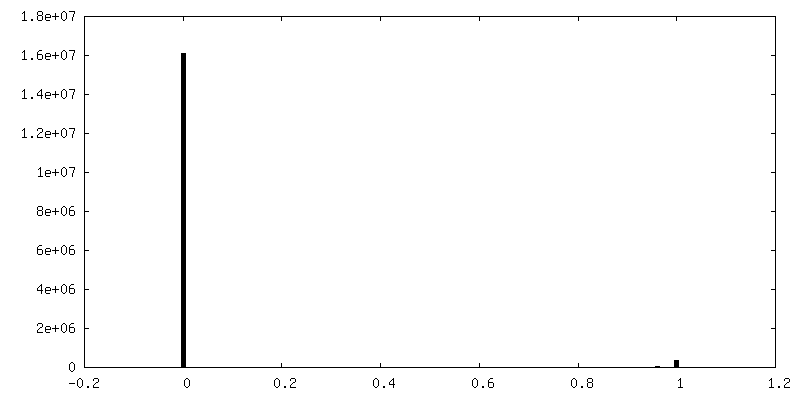
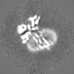
-Mask #1
| File | emd_23101_msk_1.map | ||||||||||||
|---|---|---|---|---|---|---|---|---|---|---|---|---|---|
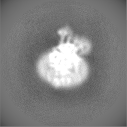
| Projections & Slices |
| ||||||||||||
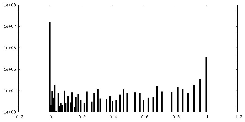
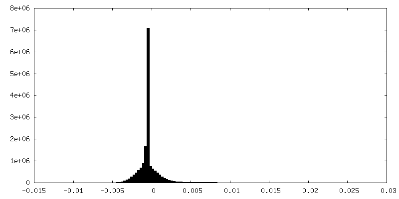
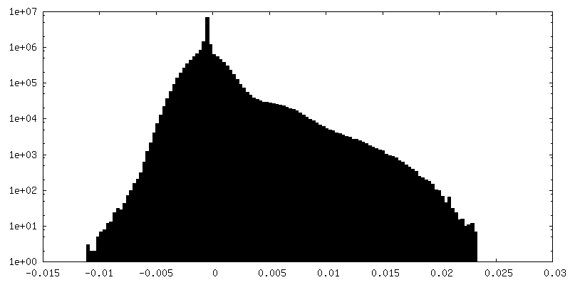
| Density Histograms |


-Half map: #1
| File | emd_23101_half_map_1.map | ||||||||||||
|---|---|---|---|---|---|---|---|---|---|---|---|---|---|
| Projections & Slices |
| ||||||||||||
| Density Histograms |


-Half map: #2
| File | emd_23101_half_map_2.map | ||||||||||||
|---|---|---|---|---|---|---|---|---|---|---|---|---|---|
| Projections & Slices |
| ||||||||||||
| Density Histograms |


- Sample components
+Entire : NTS-NTSR1-Gi complex in lipid nanodisc
+Supramolecule #1: NTS-NTSR1-Gi complex in lipid nanodisc
+Supramolecule #2: NTSR1
+Supramolecule #3: NTS
+Supramolecule #4: G(i) subunit alpha-1
+Supramolecule #5: G(I)/G(S)/G(T) subunit beta-1
+Supramolecule #6: G(T) subunit gamma-1
+Macromolecule #1: Neurotensin receptor type 1

+Macromolecule #2: Neurotensin
+Macromolecule #3: Guanine nucleotide-binding protein G(i) subunit alpha-1

 Spodoptera frugiperda (fall armyworm)
+Macromolecule #4: Guanine nucleotide-binding protein G(I)/G(S)/G(T) subunit beta-1
+Macromolecule #5: Guanine nucleotide-binding protein G(T) subunit gamma-T1
-Experimental details
-Structure determination
| Method | cryo EM |
|---|---|
 Processing Processing | single particle reconstruction |
| Aggregation state | particle |
-Sample preparation
| Buffer | pH: 6.9 |
|---|---|
| Vitrification | Cryogen name: ETHANE |
- Electron microscopy
| Microscope | FEI TITAN KRIOS |
|---|---|
| Image recording | Film or detector model: GATAN K3 BIOQUANTUM (6k x 4k) / Average electron dose: 57.0 e/Å2 |
| Electron beam | Acceleration voltage: 300 kV / Electron source:  FIELD EMISSION GUN FIELD EMISSION GUN |
| Electron optics | Illumination mode: FLOOD BEAM / Imaging mode: BRIGHT FIELD |
| Experimental equipment |  Model: Titan Krios / Image courtesy: FEI Company |
+Image processing
-Atomic model buiding 1
| Refinement | Space: REAL / Protocol: FLEXIBLE FIT |
|---|---|
| Output model | PDB-7l0r: |
