 Movie
Movie Controller

[English] 日本語
 Yorodumi
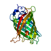
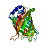
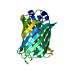
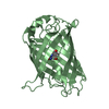
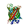
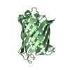
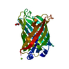
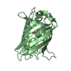
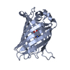
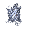
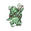
- PDB-7km4: Crystal Structure of Oxidized Version of Redox-Sensitive Superfol... -
+ Open data

- Basic information
| Entry | Database: PDB / ID: 7km4 | ||||||
|---|---|---|---|---|---|---|---|
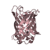
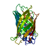
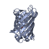
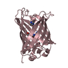
| Title | Crystal Structure of Oxidized Version of Redox-Sensitive Superfolder Green Fluorescent Protein | ||||||
 Components Components | Green fluorescent protein | ||||||
 Keywords Keywords | FLUORESCENT PROTEIN / Redox Sensitive / GFP | ||||||
| Function / homology | Green fluorescent protein, GFP / Green fluorescent protein-related / Green fluorescent protein / Green fluorescent protein / bioluminescence / generation of precursor metabolites and energy / Green fluorescent protein Function and homology information Function and homology information | ||||||
| Biological species |   Aequorea victoria (jellyfish) Aequorea victoria (jellyfish) | ||||||
| Method |  X-RAY DIFFRACTION / SYNCHROTRON / MOLECULAR REPLACEMENT / Resolution: 2.65 Å X-RAY DIFFRACTION / SYNCHROTRON / MOLECULAR REPLACEMENT / Resolution: 2.65 Å | ||||||
 Authors Authors | Nguyen, T. / Nicely, N.I. / McCafferty, D.G. | ||||||
| Funding support |  United States, 1items United States, 1items
| ||||||
 Citation Citation | Journal: To be Published Title: Crystal Structure of Oxidized Version of Redox-Sensitive Superfolder Green Fluorescent Protein Authors: Nguyen, T. / Nicely, N.I. / McCafferty, D.G. | ||||||
| History |
|
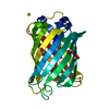
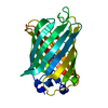
- Structure visualization
| Structure viewer | Molecule: MolmilJmol/JSmol |
|---|
- Downloads & links
-Download
| PDBx/mmCIF format | 7km4.cif.gz | 66.7 KB | Display | PDBx/mmCIF format |
|---|---|---|---|---|
| PDB format | pdb7km4.ent.gz | 40.6 KB | Display | PDB format |
| PDBx/mmJSON format | 7km4.json.gz | Tree view | PDBx/mmJSON format | |
| Others |  Other downloads Other downloads |
-Validation report
| Summary document | 7km4_validation.pdf.gz | 432.4 KB | Display | wwPDB validaton report |
|---|---|---|---|---|
| Full document | 7km4_full_validation.pdf.gz | 436.5 KB | Display | |
| Data in XML | 7km4_validation.xml.gz | 11 KB | Display | |
| Data in CIF | 7km4_validation.cif.gz | 13.7 KB | Display | |
| Arichive directory | https://data.pdbj.org/pub/pdb/validation_reports/km/7km4ftp://data.pdbj.org/pub/pdb/validation_reports/km/7km4 | HTTPS FTP |
-Related structure data
| Related structure data |  2b3pS S: Starting model for refinement |
|---|---|
| Similar structure data |

-Links
 PDBj
PDBj

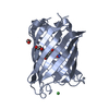
- Assembly
| Deposited unit | 
| ||||||||||||
|---|---|---|---|---|---|---|---|---|---|---|---|---|---|
| 1 |
| ||||||||||||
| Unit cell |
|
-Components
| #1: Protein | Mass: 27845.365 Da / Num. of mol.: 1 Source method: isolated from a genetically manipulated source Source: (gene. exp.) Aequorea victoria (jellyfish) / Gene: GFP / Production host:  |
|---|---|
| #2: Water | ChemComp-HOH /  Mass: 18.015 Da / Num. of mol.: 9 / Source method: isolated from a natural source / Formula: H2O Mass: 18.015 Da / Num. of mol.: 9 / Source method: isolated from a natural source / Formula: H2O |
| Has ligand of interest | Y |
| Has protein modification | Y |
-Experimental details
-Experiment
| Experiment | Method: X-RAY DIFFRACTION / Number of used crystals: 1 |
|---|
- Sample preparation
| Crystal | Density Matthews: 2.12 Å3/Da / Density % sol: 42.03 % |
|---|---|
| Crystal grow | Temperature: 298 K / Method: vapor diffusion, sitting drop / Details: 200, Potassium Chloride, 20% PEG-3350 / Temp details: Room Temperature |
-Data collection
| Diffraction | Mean temperature: 298 K / Serial crystal experiment: N |
|---|---|
| Diffraction source | Source: SYNCHROTRON / Site: APS / Beamline: 22-BM / Wavelength: 1 Å |
| Detector | Type: MARMOSAIC 225 mm CCD / Detector: CCD / Date: Aug 10, 2017 |
| Radiation | Protocol: SINGLE WAVELENGTH / Monochromatic (M) / Laue (L): M / Scattering type: x-ray |
| Radiation wavelength | Wavelength: 1 Å / Relative weight: 1 |
| Reflection | Resolution: 2.65→50 Å / Num. obs: 7433 / % possible obs: 100 % / Redundancy: 10.3 % / Biso Wilson estimate: 56.56 Å2 / Rpim(I) all: 0.031 / Rsym value: 0.059 / Net I/σ(I): 3.3 |
| Reflection shell | Resolution: 2.65→2.7 Å / Redundancy: 10.3 % / Num. unique obs: 1426 / Rpim(I) all: 0.249 / Rsym value: 0.808 / % possible all: 100 |
- Processing
| Software |
| ||||||||||||||||||||||||||||||||||||||||||
|---|---|---|---|---|---|---|---|---|---|---|---|---|---|---|---|---|---|---|---|---|---|---|---|---|---|---|---|---|---|---|---|---|---|---|---|---|---|---|---|---|---|---|---|
| Refinement | Method to determine structure: MOLECULAR REPLACEMENT Starting model: 2B3P Resolution: 2.65→40.43 Å / SU ML: 0.3792 / Cross valid method: FREE R-VALUE / σ(F): 1.36 / Phase error: 32.2407 Stereochemistry target values: GeoStd + Monomer Library + CDL v1.2
| ||||||||||||||||||||||||||||||||||||||||||
| Solvent computation | Shrinkage radii: 0.9 Å / VDW probe radii: 1.11 Å / Solvent model: FLAT BULK SOLVENT MODEL | ||||||||||||||||||||||||||||||||||||||||||
| Displacement parameters | Biso mean: 57.42 Å2 | ||||||||||||||||||||||||||||||||||||||||||
| Refinement step | Cycle: LAST / Resolution: 2.65→40.43 Å
| ||||||||||||||||||||||||||||||||||||||||||
| Refine LS restraints |
| ||||||||||||||||||||||||||||||||||||||||||
| LS refinement shell |
|
