 Movie
Movie Controller


+ Open data

- Basic information
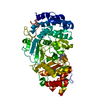
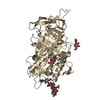
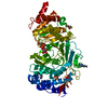
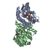
| Entry | Database: PDB / ID: 7kg1 | ||||||
|---|---|---|---|---|---|---|---|
| Title | Structure of human PARG complexed with PARG-002 | ||||||
 Components Components | Poly(ADP-ribose) glycohydrolase | ||||||
 Keywords Keywords | HYDROLASE/HYDROLASE inhibitor / HYDROLASE / hydrolase inhibitor complex / methyl xanthine / HYDROLASE-HYDROLASE inhibitor complex | ||||||
| Function / homology |  Function and homology information Function and homology informationnucleotide-sugar metabolic process / poly(ADP-ribose) glycohydrolase / poly(ADP-ribose) glycohydrolase activity / ATP generation from poly-ADP-D-ribose / POLB-Dependent Long Patch Base Excision Repair / regulation of DNA repair / base-excision repair, gap-filling / carbohydrate metabolic process / nuclear body / mitochondrial matrix ...nucleotide-sugar metabolic process / poly(ADP-ribose) glycohydrolase / poly(ADP-ribose) glycohydrolase activity / ATP generation from poly-ADP-D-ribose / POLB-Dependent Long Patch Base Excision Repair / regulation of DNA repair / base-excision repair, gap-filling / carbohydrate metabolic process / nuclear body / mitochondrial matrix / nucleoplasm / nucleus / cytosol / cytoplasm Similarity search - Function | ||||||
| Biological species |  Homo sapiens (human) Homo sapiens (human) | ||||||
| Method |  X-RAY DIFFRACTION / SYNCHROTRON / MOLECULAR REPLACEMENT / Resolution: 1.65 Å X-RAY DIFFRACTION / SYNCHROTRON / MOLECULAR REPLACEMENT / Resolution: 1.65 Å | ||||||
 Authors Authors | Brosey, C.A. / Balapiti-Modarage, L.P.F. / Warden, L.S. / Jones, D.E. / Ahmed, Z. / Tainer, J.A. | ||||||
| Funding support |  United States, 1items United States, 1items
| ||||||
 Citation Citation | Journal: Prog.Biophys.Mol.Biol. / Year: 2021 Title: Targeting SARS-CoV-2 Nsp3 macrodomain structure with insights from human poly(ADP-ribose) glycohydrolase (PARG) structures with inhibitors. Authors: Brosey, C.A. / Houl, J.H. / Katsonis, P. / Balapiti-Modarage, L.P.F. / Bommagani, S. / Arvai, A. / Moiani, D. / Bacolla, A. / Link, T. / Warden, L.S. / Lichtarge, O. / Jones, D.E. / Ahmed, Z. / Tainer, J.A. | ||||||
| History |
|
- Structure visualization
| Structure viewer | Molecule: MolmilJmol/JSmol |
|---|
- Downloads & links
-Download
| PDBx/mmCIF format | 7kg1.cif.gz | 363.5 KB | Display | PDBx/mmCIF format |
|---|---|---|---|---|
| PDB format | pdb7kg1.ent.gz | 245.4 KB | Display | PDB format |
| PDBx/mmJSON format | 7kg1.json.gz | Tree view | PDBx/mmJSON format | |
| Others |  Other downloads Other downloads |
-Validation report
| Arichive directory | https://data.pdbj.org/pub/pdb/validation_reports/kg/7kg1ftp://data.pdbj.org/pub/pdb/validation_reports/kg/7kg1 | HTTPS FTP |
|---|
-Related structure data
| Related structure data | 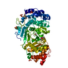 7kfpC 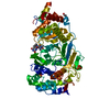 7kg0C 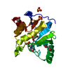 7kg3C 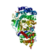 7kg6C 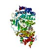 7kg7C 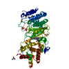 7kg8C 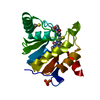 7kxbC 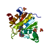 7lg7C 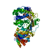 4b1gS C: citing same article ( S: Starting model for refinement |
|---|---|
| Similar structure data |

-Links
 PDBj
PDBj


- Assembly
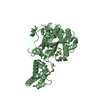
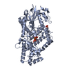
| Deposited unit | 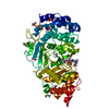
| ||||||||||
|---|---|---|---|---|---|---|---|---|---|---|---|
| 1 |
| ||||||||||
| Unit cell |
|
-Components
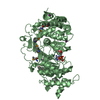
-Protein , 1 types, 1 molecules A
| #1: Protein | Mass: 61020.375 Da / Num. of mol.: 1 / Mutation: K616A, Q617A, K618A, E688A, K689A, K690A Source method: isolated from a genetically manipulated source Source: (gene. exp.) Homo sapiens (human) / Gene: PARG / Production host:  References: UniProt: Q86W56, poly(ADP-ribose) glycohydrolase |
|---|
-Non-polymers , 6 types, 316 molecules 
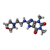


| #2: Chemical | ChemComp-DMS /  Mass: 78.133 Da / Num. of mol.: 4 / Source method: obtained synthetically / Formula: C2H6OS / Comment: DMSO, precipitant*YM Mass: 78.133 Da / Num. of mol.: 4 / Source method: obtained synthetically / Formula: C2H6OS / Comment: DMSO, precipitant*YM#3: Chemical | ChemComp-WDD / |  Mass: 308.336 Da / Num. of mol.: 1 / Source method: obtained synthetically / Formula: C13H20N6O3 / Feature type: SUBJECT OF INVESTIGATION Mass: 308.336 Da / Num. of mol.: 1 / Source method: obtained synthetically / Formula: C13H20N6O3 / Feature type: SUBJECT OF INVESTIGATION#4: Chemical |  Mass: 92.094 Da / Num. of mol.: 2 / Source method: obtained synthetically / Formula: C3H8O3 Mass: 92.094 Da / Num. of mol.: 2 / Source method: obtained synthetically / Formula: C3H8O3#5: Chemical | ChemComp-CAC / |  Mass: 136.989 Da / Num. of mol.: 1 / Source method: obtained synthetically / Formula: C2H6AsO2 Mass: 136.989 Da / Num. of mol.: 1 / Source method: obtained synthetically / Formula: C2H6AsO2#6: Chemical | ChemComp-SO4 /  Mass: 96.063 Da / Num. of mol.: 5 / Source method: obtained synthetically / Formula: SO4 Mass: 96.063 Da / Num. of mol.: 5 / Source method: obtained synthetically / Formula: SO4#7: Water | ChemComp-HOH / | Mass: 18.015 Da / Num. of mol.: 303 / Source method: isolated from a natural source / Formula: H2O |
|---|
-Details
| Has ligand of interest | Y |
|---|---|
| Has protein modification | Y |
-Experimental details
-Experiment
| Experiment | Method: X-RAY DIFFRACTION / Number of used crystals: 1 |
|---|
- Sample preparation
| Crystal | Density Matthews: 2.35 Å3/Da / Density % sol: 47.7 % |
|---|---|
| Crystal grow | Temperature: 295 K / Method: vapor diffusion, hanging drop / pH: 7.5 / Details: 0.1 M PCTP, pH 7.5, 0.2 M AmSO4, 18-23% PEG3350 |
-Data collection
| Diffraction | Mean temperature: 100 K / Serial crystal experiment: N |
|---|---|
| Diffraction source | Source: SYNCHROTRON / Site: SSRL / Beamline: BL9-2 / Wavelength: 0.9795 Å |
| Detector | Type: DECTRIS PILATUS 6M / Detector: PIXEL / Date: Feb 6, 2020 |
| Radiation | Monochromator: Si(111) and Si(220) double crystal / Protocol: SINGLE WAVELENGTH / Monochromatic (M) / Laue (L): M / Scattering type: x-ray |
| Radiation wavelength | Wavelength: 0.9795 Å / Relative weight: 1 |
| Reflection | Resolution: 1.65→38.66 Å / Num. obs: 68294 / % possible obs: 99.74 % / Redundancy: 13.1 % / Biso Wilson estimate: 26.99 Å2 / CC1/2: 1 / CC star: 1 / Rmerge(I) obs: 0.05614 / Rpim(I) all: 0.01608 / Rrim(I) all: 0.05845 / Net I/σ(I): 24.76 |
| Reflection shell | Resolution: 1.65→1.709 Å / Redundancy: 13.5 % / Rmerge(I) obs: 1.655 / Mean I/σ(I) obs: 1.74 / Num. unique obs: 6747 / CC1/2: 0.745 / CC star: 0.924 / Rpim(I) all: 0.4605 / Rrim(I) all: 1.719 / % possible all: 99.69 |
- Processing
| Software |
| |||||||||||||||||||||||||||||||||||||||||||||||||||||||||||||||||||||||||||||||||||||||||||||||||||||||||||||||||||||||||||||||||||||||||||||||||||||||||||||||||||||||||||||||
|---|---|---|---|---|---|---|---|---|---|---|---|---|---|---|---|---|---|---|---|---|---|---|---|---|---|---|---|---|---|---|---|---|---|---|---|---|---|---|---|---|---|---|---|---|---|---|---|---|---|---|---|---|---|---|---|---|---|---|---|---|---|---|---|---|---|---|---|---|---|---|---|---|---|---|---|---|---|---|---|---|---|---|---|---|---|---|---|---|---|---|---|---|---|---|---|---|---|---|---|---|---|---|---|---|---|---|---|---|---|---|---|---|---|---|---|---|---|---|---|---|---|---|---|---|---|---|---|---|---|---|---|---|---|---|---|---|---|---|---|---|---|---|---|---|---|---|---|---|---|---|---|---|---|---|---|---|---|---|---|---|---|---|---|---|---|---|---|---|---|---|---|---|---|---|---|---|
| Refinement | Method to determine structure: MOLECULAR REPLACEMENT Starting model: 4B1G Resolution: 1.65→38.66 Å / SU ML: 0.1482 / Cross valid method: FREE R-VALUE / σ(F): 1.34 / Phase error: 20.981 Stereochemistry target values: GeoStd + Monomer Library + CDL v1.2
| |||||||||||||||||||||||||||||||||||||||||||||||||||||||||||||||||||||||||||||||||||||||||||||||||||||||||||||||||||||||||||||||||||||||||||||||||||||||||||||||||||||||||||||||
| Solvent computation | Shrinkage radii: 0.9 Å / VDW probe radii: 1.11 Å / Solvent model: FLAT BULK SOLVENT MODEL | |||||||||||||||||||||||||||||||||||||||||||||||||||||||||||||||||||||||||||||||||||||||||||||||||||||||||||||||||||||||||||||||||||||||||||||||||||||||||||||||||||||||||||||||
| Displacement parameters | Biso mean: 35.24 Å2 | |||||||||||||||||||||||||||||||||||||||||||||||||||||||||||||||||||||||||||||||||||||||||||||||||||||||||||||||||||||||||||||||||||||||||||||||||||||||||||||||||||||||||||||||
| Refinement step | Cycle: LAST / Resolution: 1.65→38.66 Å
| |||||||||||||||||||||||||||||||||||||||||||||||||||||||||||||||||||||||||||||||||||||||||||||||||||||||||||||||||||||||||||||||||||||||||||||||||||||||||||||||||||||||||||||||
| Refine LS restraints |
| |||||||||||||||||||||||||||||||||||||||||||||||||||||||||||||||||||||||||||||||||||||||||||||||||||||||||||||||||||||||||||||||||||||||||||||||||||||||||||||||||||||||||||||||
| LS refinement shell |
| |||||||||||||||||||||||||||||||||||||||||||||||||||||||||||||||||||||||||||||||||||||||||||||||||||||||||||||||||||||||||||||||||||||||||||||||||||||||||||||||||||||||||||||||
| Refinement TLS params. | Method: refined / Refine-ID: X-RAY DIFFRACTION
| |||||||||||||||||||||||||||||||||||||||||||||||||||||||||||||||||||||||||||||||||||||||||||||||||||||||||||||||||||||||||||||||||||||||||||||||||||||||||||||||||||||||||||||||
| Refinement TLS group | Refine-ID: X-RAY DIFFRACTION / Auth asym-ID: A / Label asym-ID: A
|
