 Movie
Movie Controller

[English] 日本語
 Yorodumi
- PDB-4na0: Crystal structure of mouse poly(ADP-ribose) glycohydrolase (PARG)... -
+ Open data

- Basic information
| Entry | Database: PDB / ID: 4na0 | ||||||
|---|---|---|---|---|---|---|---|
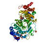
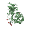
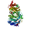
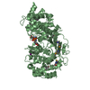
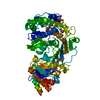
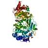
| Title | Crystal structure of mouse poly(ADP-ribose) glycohydrolase (PARG) catalytic domain with ADPRibose | ||||||
 Components Components | Poly(ADP-ribose) glycohydrolase | ||||||
 Keywords Keywords | HYDROLASE / Poly(ADP-ribose) glycohydrolase | ||||||
| Function / homology |  Function and homology information Function and homology informationPOLB-Dependent Long Patch Base Excision Repair / nucleotide-sugar metabolic process / poly(ADP-ribose) glycohydrolase / poly(ADP-ribose) glycohydrolase activity / ATP generation from poly-ADP-D-ribose / detection of bacterium / regulation of DNA repair / positive regulation of DNA repair / carbohydrate metabolic process / nuclear body ...POLB-Dependent Long Patch Base Excision Repair / nucleotide-sugar metabolic process / poly(ADP-ribose) glycohydrolase / poly(ADP-ribose) glycohydrolase activity / ATP generation from poly-ADP-D-ribose / detection of bacterium / regulation of DNA repair / positive regulation of DNA repair / carbohydrate metabolic process / nuclear body / chromatin binding / DNA damage response / chromatin / nucleoplasm / nucleus / cytoplasm / cytosol Similarity search - Function | ||||||
| Biological species |  | ||||||
| Method |  X-RAY DIFFRACTION / SYNCHROTRON / molecular replacement-SAD / Resolution: 2.4 Å X-RAY DIFFRACTION / SYNCHROTRON / molecular replacement-SAD / Resolution: 2.4 Å | ||||||
 Authors Authors | Wang, Z. / Cheng, Z. / Xu, W. | ||||||
 Citation Citation | Journal: Plos One / Year: 2014 Title: Crystallographic and biochemical analysis of the mouse poly(ADP-ribose) glycohydrolase. Authors: Wang, Z. / Gagne, J.P. / Poirier, G.G. / Xu, W. | ||||||
| History |
|
- Structure visualization
| Structure viewer | Molecule: MolmilJmol/JSmol |
|---|
- Downloads & links
-Download
| PDBx/mmCIF format | 4na0.cif.gz | 633.2 KB | Display | PDBx/mmCIF format |
|---|---|---|---|---|
| PDB format | pdb4na0.ent.gz | 527.3 KB | Display | PDB format |
| PDBx/mmJSON format | 4na0.json.gz | Tree view | PDBx/mmJSON format | |
| Others |  Other downloads Other downloads |
-Validation report
| Arichive directory | https://data.pdbj.org/pub/pdb/validation_reports/na/4na0ftp://data.pdbj.org/pub/pdb/validation_reports/na/4na0 | HTTPS FTP |
|---|
-Related structure data
| Related structure data |  4fc2C  4n9yC  4n9zC  4na4C  4na5C  4na6C C: citing same article ( |
|---|---|
| Similar structure data |


-Links
 PDBj
PDBj
- Assembly
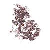
| Deposited unit | 
| ||||||||
|---|---|---|---|---|---|---|---|---|---|
| 1 | 
| ||||||||
| 2 | 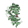
| ||||||||
| 3 | 
| ||||||||
| Unit cell |
|
-Components
| #1: Protein | Mass: 60569.629 Da / Num. of mol.: 3 / Fragment: catalytic domain Source method: isolated from a genetically manipulated source Source: (gene. exp.)  References: UniProt: O88622, poly(ADP-ribose) glycohydrolase #2: Chemical | ChemComp-IOD /   Mass: 126.904 Da / Num. of mol.: 6 / Source method: obtained synthetically / Formula: I Mass: 126.904 Da / Num. of mol.: 6 / Source method: obtained synthetically / Formula: I#3: Chemical |   Mass: 559.316 Da / Num. of mol.: 3 / Source method: obtained synthetically / Formula: C15H23N5O14P2 Mass: 559.316 Da / Num. of mol.: 3 / Source method: obtained synthetically / Formula: C15H23N5O14P2Has protein modification | Y | |
|---|
-Experimental details
-Experiment
| Experiment | Method: X-RAY DIFFRACTION / Number of used crystals: 1 |
|---|
- Sample preparation
| Crystal | Density Matthews: 2.43 Å3/Da / Density % sol: 49.39 % |
|---|---|
| Crystal grow | Temperature: 295 K / Method: vapor diffusion, hanging drop / pH: 7 Details: 0.22M KI 20% PEG3,350, pH 7, VAPOR DIFFUSION, HANGING DROP, temperature 295K |
-Data collection
| Diffraction | Mean temperature: 100 K |
|---|---|
| Diffraction source | Source: SYNCHROTRON / Site: ALS  / Beamline: 8.2.1 / Wavelength: 0.9774 Å / Beamline: 8.2.1 / Wavelength: 0.9774 Å |
| Detector | Type: ADSC QUANTUM 315r / Detector: CCD / Date: Dec 5, 2011 |
| Radiation | Monochromator: Double crystal, Si(111) / Protocol: SINGLE WAVELENGTH / Monochromatic (M) / Laue (L): M / Scattering type: x-ray |
| Radiation wavelength | Wavelength: 0.9774 Å / Relative weight: 1 |
| Reflection | Resolution: 2.4→50 Å / Num. all: 68770 / Num. obs: 67670 / % possible obs: 98.4 % / Observed criterion σ(F): 2 / Observed criterion σ(I): 2 / Rmerge(I) obs: 0.113 |
| Reflection shell | Resolution: 2.4→2.47 Å |
- Processing
| Software |
| ||||||||||||||||||||||||||||||||||||||||||||||||||||||||||||||||||||||||||||||||||||||||||||||||||||
|---|---|---|---|---|---|---|---|---|---|---|---|---|---|---|---|---|---|---|---|---|---|---|---|---|---|---|---|---|---|---|---|---|---|---|---|---|---|---|---|---|---|---|---|---|---|---|---|---|---|---|---|---|---|---|---|---|---|---|---|---|---|---|---|---|---|---|---|---|---|---|---|---|---|---|---|---|---|---|---|---|---|---|---|---|---|---|---|---|---|---|---|---|---|---|---|---|---|---|---|---|---|
| Refinement | Method to determine structure: molecular replacement-SAD / Resolution: 2.4→50 Å / Cor.coef. Fo:Fc: 0.923 / Cor.coef. Fo:Fc free: 0.901 / Occupancy max: 1 / Occupancy min: 1 / SU B: 34.856 / SU ML: 0.379 / Cross valid method: THROUGHOUT / σ(F): 0 / ESU R: 0.673 / ESU R Free: 0.37 / Stereochemistry target values: MAXIMUM LIKELIHOOD Details: HYDROGENS HAVE BEEN ADDED IN THE RIDING POSITIONS U VALUES : WITH TLS ADDED
| ||||||||||||||||||||||||||||||||||||||||||||||||||||||||||||||||||||||||||||||||||||||||||||||||||||
| Solvent computation | Ion probe radii: 0.8 Å / Shrinkage radii: 0.8 Å / VDW probe radii: 1.4 Å / Solvent model: BABINET MODEL WITH MASK | ||||||||||||||||||||||||||||||||||||||||||||||||||||||||||||||||||||||||||||||||||||||||||||||||||||
| Displacement parameters | Biso max: 290.86 Å2 / Biso mean: 70.4119 Å2 / Biso min: 16.68 Å2
| ||||||||||||||||||||||||||||||||||||||||||||||||||||||||||||||||||||||||||||||||||||||||||||||||||||
| Refinement step | Cycle: LAST / Resolution: 2.4→50 Å
| ||||||||||||||||||||||||||||||||||||||||||||||||||||||||||||||||||||||||||||||||||||||||||||||||||||
| Refine LS restraints |
| ||||||||||||||||||||||||||||||||||||||||||||||||||||||||||||||||||||||||||||||||||||||||||||||||||||
| LS refinement shell | Resolution: 2.404→2.467 Å / Total num. of bins used: 20
| ||||||||||||||||||||||||||||||||||||||||||||||||||||||||||||||||||||||||||||||||||||||||||||||||||||
| Refinement TLS params. | Method: refined / Refine-ID: X-RAY DIFFRACTION
| ||||||||||||||||||||||||||||||||||||||||||||||||||||||||||||||||||||||||||||||||||||||||||||||||||||
| Refinement TLS group |
|
