- PDB-4b1h: Structure of human PARG catalytic domain in complex with ADP-ribose -
+
Open data
ID or keywords:
Loading...
-
Basic information
Entry
Database: PDB / ID: 4b1h
Title
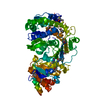
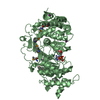
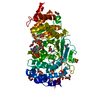
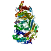
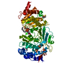
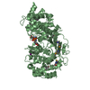
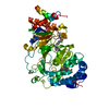
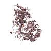
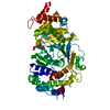
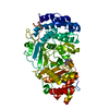
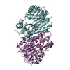
Structure of human PARG catalytic domain in complex with ADP-ribose
Components
POLY(ADP-RIBOSE) GLYCOHYDROLASE
Keywords
HYDROLASE
Function / homology
Function and homology information
nucleotide-sugar metabolic process / poly(ADP-ribose) glycohydrolase / poly(ADP-ribose) glycohydrolase activity / ATP generation from poly-ADP-D-ribose / POLB-Dependent Long Patch Base Excision Repair / regulation of DNA repair / base-excision repair, gap-filling / carbohydrate metabolic process / nuclear body / mitochondrial matrix ...nucleotide-sugar metabolic process / poly(ADP-ribose) glycohydrolase / poly(ADP-ribose) glycohydrolase activity / ATP generation from poly-ADP-D-ribose / POLB-Dependent Long Patch Base Excision Repair / regulation of DNA repair / base-excision repair, gap-filling / carbohydrate metabolic process / nuclear body / mitochondrial matrix / nucleoplasm / nucleus / cytoplasm / cytosol Similarity search - Function
Mass: 18.015 Da / Num. of mol.: 378 / Source method: isolated from a natural source / Formula: H2O
-
Details
Nonpolymer details
DTV: DTT FROM PROTEIN BUFFER DERIVATISES CYSTEINE TO FORM (2R)-2-AZANIUMYL-3-[[(2S,3S)-2,3- ...DTV: DTT FROM PROTEIN BUFFER DERIVATISES CYSTEINE TO FORM (2R)-2-AZANIUMYL-3-[[(2S,3S)-2,3-DIHYDROXY-4-SULFANYL] PROPANOATE
Sequence details
CODON OPTIMISED SEQUENCE, SIX MUTATIONS INTRODUCED TO ENHANCE CRYSTALLISABILITY ...CODON OPTIMISED SEQUENCE, SIX MUTATIONS INTRODUCED TO ENHANCE CRYSTALLISABILITY K617A,Q618A,K619A,E688A,K689A, K690A
-
Experimental details
-
Experiment
Experiment
Method: X-RAY DIFFRACTION / Number of used crystals: 1
-
Sample preparation
Crystal
Density Matthews: 2.48 Å3/Da / Density % sol: 50.47 % / Description: NONE
Resolution: 2→27.35 Å / Cor.coef. Fo:Fc: 0.955 / Cor.coef. Fo:Fc free: 0.94 / SU B: 8.39 / SU ML: 0.1 / Cross valid method: THROUGHOUT / ESU R: 0.2 / ESU R Free: 0.17 / Stereochemistry target values: MAXIMUM LIKELIHOOD / Details: HYDROGENS HAVE BEEN ADDED IN THE RIDING POSITIONS.
Rfactor
Num. reflection
% reflection
Selection details
Rfree
0.24139
1961
5 %
RANDOM
Rwork
0.20356
-
-
-
obs
0.20548
37234
98.62 %
-
Solvent computation
Ion probe radii: 0.8 Å / Shrinkage radii: 0.8 Å / VDW probe radii: 1.4 Å / Solvent model: BABINET MODEL WITH MASK
 Movie
Movie Controller

 Yorodumi
 Open data
 Basic information
 Components
 Keywords
 Function and homology information
 HOMO SAPIENS (human)
 X-RAY DIFFRACTION /
  Authors
 Citation
 Structure visualization
 Downloads & links
 Other downloads


 PDBj
PDBj


 Assembly
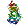


 Mass: 78.133 Da / Num. of mol.: 1 / Source method: obtained synthetically / Formula: C2H6OS
 Mass: 154.251 Da / Num. of mol.: 1 / Source method: obtained synthetically / Formula: C4H10O2S2
 Mass: 92.094 Da / Num. of mol.: 2 / Source method: obtained synthetically / Formula: C3H8O3
 Mass: 559.316 Da / Num. of mol.: 2 / Source method: obtained synthetically / Formula: C15H23N5O14P2
 Mass: 96.063 Da / Num. of mol.: 2 / Source method: obtained synthetically / Formula: SO4
 Sample preparation
 Processing