- PDB-6hmk: POLYADPRIBOSYL GLYCOHYDROLASE IN COMPLEX WITH PDD00016690 -
+
Open data
ID or keywords:
Loading...
-
Basic information
Entry
Database: PDB / ID: 6hmk
Title
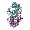
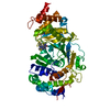
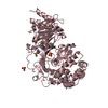
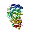
POLYADPRIBOSYL GLYCOHYDROLASE IN COMPLEX WITH PDD00016690
Components
Poly(ADP-ribose) glycohydrolase
Keywords
HYDROLASE / COMPETITIVE INHIBITOR / PARG
Function / homology
Function and homology information
nucleotide-sugar metabolic process / poly(ADP-ribose) glycohydrolase / poly(ADP-ribose) glycohydrolase activity / ATP generation from poly-ADP-D-ribose / POLB-Dependent Long Patch Base Excision Repair / regulation of DNA repair / base-excision repair, gap-filling / carbohydrate metabolic process / nuclear body / mitochondrial matrix ...nucleotide-sugar metabolic process / poly(ADP-ribose) glycohydrolase / poly(ADP-ribose) glycohydrolase activity / ATP generation from poly-ADP-D-ribose / POLB-Dependent Long Patch Base Excision Repair / regulation of DNA repair / base-excision repair, gap-filling / carbohydrate metabolic process / nuclear body / mitochondrial matrix / nucleoplasm / nucleus / cytoplasm / cytosol Similarity search - Function
Mass: 18.015 Da / Num. of mol.: 448 / Source method: isolated from a natural source / Formula: H2O
-
Experimental details
-
Experiment
Experiment
Method: X-RAY DIFFRACTION / Number of used crystals: 1
-
Sample preparation
Crystal
Density Matthews: 2.47 Å3/Da / Density % sol: 50.22 %
Crystal grow
Temperature: 293 K / Method: vapor diffusion, sitting drop / pH: 7.5 Details: 750 nL purified protein at 7.5 mg/mL in 50 mM HEPES, pH 7.0, 150 mM NaCl, 2 mM DTT was mixed with 250 nL of seed stock and 1000 nL of a precipitant consisting of 18-23 % (w/v) PEG-3350, 0.2 ...Details: 750 nL purified protein at 7.5 mg/mL in 50 mM HEPES, pH 7.0, 150 mM NaCl, 2 mM DTT was mixed with 250 nL of seed stock and 1000 nL of a precipitant consisting of 18-23 % (w/v) PEG-3350, 0.2 M ammonium sulphate, 0.1 M PCTP pH 7.5. Seed stock was prepared using a Seed BeadTM (Hampton Research) from a co-crystal of GS-PARG(448-976 [K617A, Q618A, K619A, E688A, K689A, K690A]) with ADP-ribose, with co-crystallisation mother liquor (19 % (w/v) PEG-3350, 0.2 M ammonium sulphate, 0.1 M PCTP pH 7.5) as the stabilising solution. The final volume of the seed stock was 100 microL.
Resolution: 2.06→28.77 Å / SU R Cruickshank DPI: 0.16 / Cross valid method: THROUGHOUT / SU R Blow DPI: 0.175 / SU Rfree Blow DPI: 0.143 / SU Rfree Cruickshank DPI: 0.143
Rfactor
Num. reflection
% reflection
Selection details
Rfree
0.1888
1823
5.01 %
RANDOM
Rwork
0.1536
-
-
-
obs
0.1554
36386
99.7 %
-
Displacement parameters
Biso mean: 33.03 Å2
Baniso -1
Baniso -2
Baniso -3
1-
6.2142 Å2
0 Å2
0 Å2
2-
-
3.0208 Å2
0 Å2
3-
-
-
-9.235 Å2
Refine analyze
Luzzati coordinate error obs: 0.211 Å
Refinement step
Cycle: LAST / Resolution: 2.06→28.77 Å
Protein
Nucleic acid
Ligand
Solvent
Total
Num. atoms
4028
0
62
448
4538
Refine LS restraints
Refine-ID
Type
Dev ideal
Number
Restraint function
Weight
X-RAY DIFFRACTION
BONDANGLES
0.96
5850
HARMONIC
2
X-RAY DIFFRACTION
BONDLENGTHS
0.01
4286
HARMONIC
2
X-RAY DIFFRACTION
PEPTIDEOMEGATORSIONANGLES
3.43
1458
SINUSOIDAL
2
X-RAY DIFFRACTION
OTHERTORSIONANGLES
16.45
SINUSOIDAL
2
LS refinement shell
Resolution: 2.06→2.12 Å
Rfactor
Num. reflection
% reflection
Rfree
0.2213
130
4.42 %
Rwork
0.1862
2809
-
obs
-
-
99.7 %
+
About Yorodumi
-
News
-
Feb 9, 2022. New format data for meta-information of EMDB entries
New format data for meta-information of EMDB entries
Version 3 of the EMDB header file is now the official format.
The previous official version 1.9 will be removed from the archive.
In the structure databanks used in Yorodumi, some data are registered as the other names, "COVID-19 virus" and "2019-nCoV". Here are the details of the virus and the list of structure data.
Jan 31, 2019. EMDB accession codes are about to change! (news from PDBe EMDB page)
EMDB accession codes are about to change! (news from PDBe EMDB page)
The allocation of 4 digits for EMDB accession codes will soon come to an end. Whilst these codes will remain in use, new EMDB accession codes will include an additional digit and will expand incrementally as the available range of codes is exhausted. The current 4-digit format prefixed with “EMD-” (i.e. EMD-XXXX) will advance to a 5-digit format (i.e. EMD-XXXXX), and so on. It is currently estimated that the 4-digit codes will be depleted around Spring 2019, at which point the 5-digit format will come into force.
The EM Navigator/Yorodumi systems omit the EMD- prefix.
Related info.:Q: What is EMD? / ID/Accession-code notation in Yorodumi/EM Navigator
Yorodumi is a browser for structure data from EMDB, PDB, SASBDB, etc.
This page is also the successor to EM Navigator detail page, and also detail information page/front-end page for Omokage search.
The word "yorodu" (or yorozu) is an old Japanese word meaning "ten thousand". "mi" (miru) is to see.
Related info.:EMDB / PDB / SASBDB / Comparison of 3 databanks / Yorodumi Search / Aug 31, 2016. New EM Navigator & Yorodumi / Yorodumi Papers / Jmol/JSmol / Function and homology information / Changes in new EM Navigator and Yorodumi
 Movie
Movie Controller


 Open data
 Basic information
 Components
 Keywords
 Function and homology information
 Homo sapiens (human)
 X-RAY DIFFRACTION /
  Authors
 Citation
 Structure visualization
 Downloads & links
 Other downloads


 PDBj
PDBj

 Assembly
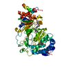


 Mass: 78.133 Da / Num. of mol.: 2 / Source method: obtained synthetically / Formula: C2H6OS / Comment: DMSO, precipitant*YM
 Mass: 92.094 Da / Num. of mol.: 1 / Source method: obtained synthetically / Formula: C3H8O3
 Mass: 96.063 Da / Num. of mol.: 4 / Source method: obtained synthetically / Formula: SO4
 Mass: 420.506 Da / Num. of mol.: 1 / Source method: obtained synthetically / Formula: C18H20N4O4S2 / Feature type: SUBJECT OF INVESTIGATION
 Sample preparation
 / Beamline: I02 / Wavelength: 0.979 Å
 Processing