- PDB-6hmn: POLYADPRIBOSYL GLYCOSIDASE IN COMPLEX WITH PDD00014909 -
+
Open data
ID or keywords:
Loading...
-
Basic information
Entry
Database: PDB / ID: 6hmn
Title
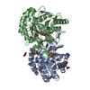
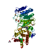
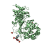
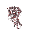
POLYADPRIBOSYL GLYCOSIDASE IN COMPLEX WITH PDD00014909
Components
Poly(ADP-ribose) glycohydrolase
Keywords
HYDROLASE / COMPETITIVE INHIBITOR
Function / homology
Function and homology information
nucleotide-sugar metabolic process / poly(ADP-ribose) glycohydrolase / poly(ADP-ribose) glycohydrolase activity / ATP generation from poly-ADP-D-ribose / POLB-Dependent Long Patch Base Excision Repair / regulation of DNA repair / base-excision repair, gap-filling / carbohydrate metabolic process / nuclear body / mitochondrial matrix ...nucleotide-sugar metabolic process / poly(ADP-ribose) glycohydrolase / poly(ADP-ribose) glycohydrolase activity / ATP generation from poly-ADP-D-ribose / POLB-Dependent Long Patch Base Excision Repair / regulation of DNA repair / base-excision repair, gap-filling / carbohydrate metabolic process / nuclear body / mitochondrial matrix / nucleoplasm / nucleus / cytoplasm / cytosol Similarity search - Function
Mass: 18.015 Da / Num. of mol.: 102 / Source method: isolated from a natural source / Formula: H2O
-
Experimental details
-
Experiment
Experiment
Method: X-RAY DIFFRACTION / Number of used crystals: 1
-
Sample preparation
Crystal
Density Matthews: 2.49 Å3/Da / Density % sol: 50.63 %
Crystal grow
Temperature: 293 K / Method: vapor diffusion, sitting drop / pH: 7.5 Details: Protein at 7.5 mg/mL in 50 mM HEPES, pH 7.0, 150 mM NaCl, 2 mM DTT was mixed with precipitant consisting of 18 - 23 percent PEG-3350, 0.2 M ammonium sulphate, 0.1 M PCTP pH 7.5 in a 1:1 ...Details: Protein at 7.5 mg/mL in 50 mM HEPES, pH 7.0, 150 mM NaCl, 2 mM DTT was mixed with precipitant consisting of 18 - 23 percent PEG-3350, 0.2 M ammonium sulphate, 0.1 M PCTP pH 7.5 in a 1:1 ratio to give a 4 microL drop
In the structure databanks used in Yorodumi, some data are registered as the other names, "COVID-19 virus" and "2019-nCoV". Here are the details of the virus and the list of structure data.
Jan 31, 2019. EMDB accession codes are about to change! (news from PDBe EMDB page)
EMDB accession codes are about to change! (news from PDBe EMDB page)
The allocation of 4 digits for EMDB accession codes will soon come to an end. Whilst these codes will remain in use, new EMDB accession codes will include an additional digit and will expand incrementally as the available range of codes is exhausted. The current 4-digit format prefixed with “EMD-” (i.e. EMD-XXXX) will advance to a 5-digit format (i.e. EMD-XXXXX), and so on. It is currently estimated that the 4-digit codes will be depleted around Spring 2019, at which point the 5-digit format will come into force.
The EM Navigator/Yorodumi systems omit the EMD- prefix.
Related info.:Q: What is EMD? / ID/Accession-code notation in Yorodumi/EM Navigator
Yorodumi is a browser for structure data from EMDB, PDB, SASBDB, etc.
This page is also the successor to EM Navigator detail page, and also detail information page/front-end page for Omokage search.
The word "yorodu" (or yorozu) is an old Japanese word meaning "ten thousand". "mi" (miru) is to see.
Related info.:EMDB / PDB / SASBDB / Comparison of 3 databanks / Yorodumi Search / Aug 31, 2016. New EM Navigator & Yorodumi / Yorodumi Papers / Jmol/JSmol / Function and homology information / Changes in new EM Navigator and Yorodumi
 Movie
Movie Controller


 Open data
 Basic information
 Components
 Keywords
 Function and homology information
 Homo sapiens (human)
 X-RAY DIFFRACTION /
  Authors
 Citation
 Structure visualization
 Downloads & links
 Other downloads


 PDBj
PDBj

 Assembly
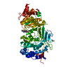


 Mass: 78.133 Da / Num. of mol.: 1 / Source method: obtained synthetically / Formula: C2H6OS / Comment: DMSO, precipitant*YM
 Mass: 92.094 Da / Num. of mol.: 1 / Source method: obtained synthetically / Formula: C3H8O3
 Mass: 401.479 Da / Num. of mol.: 1 / Source method: obtained synthetically / Formula: C20H23N3O4S / Feature type: SUBJECT OF INVESTIGATION
 Mass: 96.063 Da / Num. of mol.: 2 / Source method: obtained synthetically / Formula: SO4
 Sample preparation
 / Beamline: I04-1 / Wavelength: 0.92 Å
 Processing