 Movie
Movie Controller


+ Open data

- Basic information
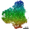
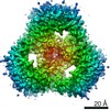
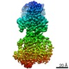
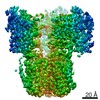
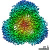
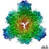
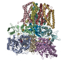
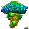
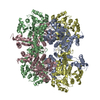
| Entry | Database: PDB / ID: 7jz2 | ||||||
|---|---|---|---|---|---|---|---|
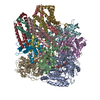
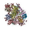
| Title | Succinate: quinone oxidoreductase SQR from E.coli K12 | ||||||
 Components Components | (Succinate dehydrogenase ...) x 4 | ||||||
 Keywords Keywords | ELECTRON TRANSPORT/OXIDOREDUCTASE / Complex / Succinate: quinone oxidoreductase / SdhA / sdhB / SdhC / SdhD / ELECTRON TRANSPORT / ELECTRON TRANSPORT-OXIDOREDUCTASE complex | ||||||
| Function / homology |  Function and homology information Function and homology informationsuccinate dehydrogenase activity / respiratory chain complex II (succinate dehydrogenase) / succinate dehydrogenase (quinone) activity / succinate dehydrogenase / cytochrome complex assembly / aerobic electron transport chain / anaerobic respiration / 3 iron, 4 sulfur cluster binding / ubiquinone binding / iron-sulfur cluster binding ...succinate dehydrogenase activity / respiratory chain complex II (succinate dehydrogenase) / succinate dehydrogenase (quinone) activity / succinate dehydrogenase / cytochrome complex assembly / aerobic electron transport chain / anaerobic respiration / 3 iron, 4 sulfur cluster binding / ubiquinone binding / iron-sulfur cluster binding / membrane => GO:0016020 / tricarboxylic acid cycle / aerobic respiration / respiratory electron transport chain / 2 iron, 2 sulfur cluster binding / flavin adenine dinucleotide binding / 4 iron, 4 sulfur cluster binding / electron transfer activity / heme binding / metal ion binding / membrane / plasma membrane Similarity search - Function | ||||||
| Biological species |  | ||||||
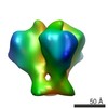
| Method | ELECTRON MICROSCOPY / single particle reconstruction / cryo EM / Resolution: 2.5 Å | ||||||
 Authors Authors | Lyu, M. / Su, C.-C. / Morgan, C.E. / Bolla, J.R. / Robinson, C.V. / Yu, E.W. | ||||||
| Funding support |  United States, 1items United States, 1items
| ||||||
 Citation Citation | Journal: Nat Methods / Year: 2021 Title: A 'Build and Retrieve' methodology to simultaneously solve cryo-EM structures of membrane proteins. Authors: Chih-Chia Su / Meinan Lyu / Christopher E Morgan / Jani Reddy Bolla / Carol V Robinson / Edward W Yu /  Abstract: Single-particle cryo-electron microscopy (cryo-EM) has become a powerful technique in the field of structural biology. However, the inability to reliably produce pure, homogeneous membrane protein ...Single-particle cryo-electron microscopy (cryo-EM) has become a powerful technique in the field of structural biology. However, the inability to reliably produce pure, homogeneous membrane protein samples hampers the progress of their structural determination. Here, we develop a bottom-up iterative method, Build and Retrieve (BaR), that enables the identification and determination of cryo-EM structures of a variety of inner and outer membrane proteins, including membrane protein complexes of different sizes and dimensions, from a heterogeneous, impure protein sample. We also use the BaR methodology to elucidate structural information from Escherichia coli K12 crude membrane and raw lysate. The findings demonstrate that it is possible to solve high-resolution structures of a number of relatively small (<100 kDa) and less abundant (<10%) unidentified membrane proteins within a single, heterogeneous sample. Importantly, these results highlight the potential of cryo-EM for systems structural proteomics. | ||||||
| History |
|
- Structure visualization
| Movie |
Movie viewer |
|---|---|
| Structure viewer | Molecule: MolmilJmol/JSmol |
- Downloads & links
-Download
| PDBx/mmCIF format | 7jz2.cif.gz | 511.6 KB | Display | PDBx/mmCIF format |
|---|---|---|---|---|
| PDB format | pdb7jz2.ent.gz | 414.8 KB | Display | PDB format |
| PDBx/mmJSON format | 7jz2.json.gz | Tree view | PDBx/mmJSON format | |
| Others |  Other downloads Other downloads |
-Validation report
| Summary document | 7jz2_validation.pdf.gz | 1.7 MB | Display | wwPDB validaton report |
|---|---|---|---|---|
| Full document | 7jz2_full_validation.pdf.gz | 1.9 MB | Display | |
| Data in XML | 7jz2_validation.xml.gz | 99.7 KB | Display | |
| Data in CIF | 7jz2_validation.cif.gz | 139.5 KB | Display | |
| Arichive directory | https://data.pdbj.org/pub/pdb/validation_reports/jz/7jz2ftp://data.pdbj.org/pub/pdb/validation_reports/jz/7jz2 | HTTPS FTP |
-Related structure data
| Related structure data | 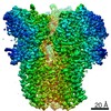 22528MC 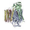 6wtiC  6wtzC 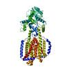 6wu0C 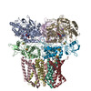 6wu6C 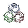 7jz3C 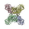 7jz6C 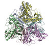 7jzhC M: map data used to model this data C: citing same article ( |
|---|---|
| Similar structure data |


-Links
 PDBj
PDBj


- Assembly
| Deposited unit | 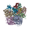
|
|---|---|
| 1 |
|
-Components
-Succinate dehydrogenase ... , 4 types, 12 molecules AEIBFJCGKDHL
| #1: Protein | Mass: 64502.766 Da / Num. of mol.: 3 / Source method: isolated from a natural source / Source: (natural) #2: Protein | Mass: 26800.912 Da / Num. of mol.: 3 / Source method: isolated from a natural source / Source: (natural) #3: Protein | Mass: 14313.100 Da / Num. of mol.: 3 / Source method: isolated from a natural source / Source: (natural) #4: Protein | Mass: 12874.438 Da / Num. of mol.: 3 / Source method: isolated from a natural source / Source: (natural) |
|---|
-Non-polymers , 8 types, 24 molecules 


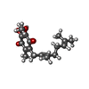
| #5: Chemical |  Mass: 785.550 Da / Num. of mol.: 3 / Source method: obtained synthetically / Formula: C27H33N9O15P2 / Comment: FAD*YM Mass: 785.550 Da / Num. of mol.: 3 / Source method: obtained synthetically / Formula: C27H33N9O15P2 / Comment: FAD*YM#6: Chemical |  Mass: 22.990 Da / Num. of mol.: 3 / Source method: obtained synthetically / Formula: Na Mass: 22.990 Da / Num. of mol.: 3 / Source method: obtained synthetically / Formula: Na#7: Chemical |  Mass: 175.820 Da / Num. of mol.: 3 / Source method: obtained synthetically / Formula: Fe2S2 Mass: 175.820 Da / Num. of mol.: 3 / Source method: obtained synthetically / Formula: Fe2S2#8: Chemical |  Mass: 351.640 Da / Num. of mol.: 3 / Source method: obtained synthetically / Formula: Fe4S4 Mass: 351.640 Da / Num. of mol.: 3 / Source method: obtained synthetically / Formula: Fe4S4#9: Chemical |  Mass: 295.795 Da / Num. of mol.: 3 / Source method: obtained synthetically / Formula: Fe3S4 / Feature type: SUBJECT OF INVESTIGATION Mass: 295.795 Da / Num. of mol.: 3 / Source method: obtained synthetically / Formula: Fe3S4 / Feature type: SUBJECT OF INVESTIGATION#10: Chemical |  Mass: 748.065 Da / Num. of mol.: 3 / Source method: obtained synthetically / Formula: C41H82NO8P / Comment: phospholipid*YM Mass: 748.065 Da / Num. of mol.: 3 / Source method: obtained synthetically / Formula: C41H82NO8P / Comment: phospholipid*YM#11: Chemical |  Mass: 616.487 Da / Num. of mol.: 3 / Source method: isolated from a natural source / Formula: C34H32FeN4O4 Mass: 616.487 Da / Num. of mol.: 3 / Source method: isolated from a natural source / Formula: C34H32FeN4O4#12: Chemical |  Mass: 318.407 Da / Num. of mol.: 3 / Source method: obtained synthetically / Formula: C19H26O4 Mass: 318.407 Da / Num. of mol.: 3 / Source method: obtained synthetically / Formula: C19H26O4 |
|---|
-Details
| Has ligand of interest | Y |
|---|
-Experimental details
-Experiment
| Experiment | Method: ELECTRON MICROSCOPY |
|---|---|
| EM experiment | Aggregation state: PARTICLE / 3D reconstruction method: single particle reconstruction |
- Sample preparation
| Component | Name: Succinate: quinone oxidoreductase SQR from E.coli K12 / Type: COMPLEX / Entity ID: #1-#4 / Source: NATURAL |
|---|---|
| Source (natural) | Organism: |
| Buffer solution | pH: 7.5 |
| Specimen | Embedding applied: NO / Shadowing applied: NO / Staining applied: NO / Vitrification applied: YES |
| Vitrification | Cryogen name: ETHANE |
- Electron microscopy imaging
| Experimental equipment |  Model: Titan Krios / Image courtesy: FEI Company |
|---|---|
| Microscopy | Model: FEI TITAN KRIOS |
| Electron gun | Electron source:  FIELD EMISSION GUN / Accelerating voltage: 300 kV / Illumination mode: FLOOD BEAM FIELD EMISSION GUN / Accelerating voltage: 300 kV / Illumination mode: FLOOD BEAM |
| Electron lens | Mode: BRIGHT FIELD |
| Image recording | Electron dose: 40 e/Å2 / Film or detector model: GATAN K3 BIOQUANTUM (6k x 4k) |
- Processing
| CTF correction | Type: PHASE FLIPPING AND AMPLITUDE CORRECTION |
|---|---|
| 3D reconstruction | Resolution: 2.5 Å / Resolution method: FSC 0.143 CUT-OFF / Num. of particles: 38471 / Symmetry type: POINT |
